Prognostic significance of histologic phenotype in periampullary adenocarcinomas
Hee-Sung Kim, Chang-Min Heo, Yoo-Shin Choi, Suk-Won Suh, Seung Eun Lee

TL;DR
This study finds that the histologic type of periampullary adenocarcinomas, specifically intestinal versus pancreatobiliary, is linked to different survival rates and tumor behaviors.
Contribution
The study identifies clinicopathological differences and survival outcomes between intestinal and pancreatobiliary types of periampullary adenocarcinomas.
Findings
Intestinal-type tumors were associated with better survival and less aggressive features compared to pancreatobiliary-type tumors.
The 5-year overall survival rate was significantly higher for intestinal-type tumors (58.8%) than for pancreatobiliary-type tumors (20.4%).
Curative resection was the only independent prognostic factor in multivariate analysis.
Abstract
Periampullary adenocarcinomas typically exhibit either intestinal or pancreatobiliary (PB) differentiation, and the type of differentiation may be prognostically more important than the anatomic site of origin. This study aimed to evaluate prognostic significance of histological type of periampullary carcinomas. Microscopic slides from 110 consecutive pancreatoduodenectomies performed between 2010 and 2020 were reviewed and classified as intestinal or PB type. Clinicopathological factors were compared between PB-(n=93) and intestinal-type (n=17) differentiation. The intestinal type included significantly more patients with well-differentiated histology (35.3% vs. 11.8%, p=0.001) and fewer patients with perineural invasion (41.2% vs. 76.4%, p=0.029), advanced T stage (> T3; 41.2% vs.74.2%, p=0.007), and systemic recurrence (71.4% vs. 92.9%, p=0.005) than PB type. The 5-year-overall…
Genes, proteins, chemicals, diseases, species, mutations and cell lines named across the full text — each resolved to its canonical identifier and authoritative record.
Click any figure to enlarge with its caption.
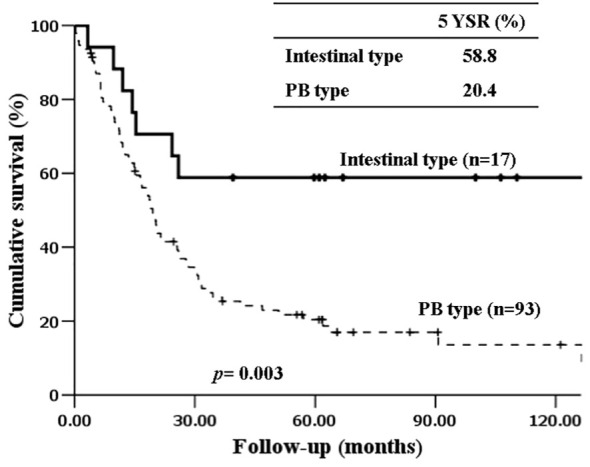 Figure 1
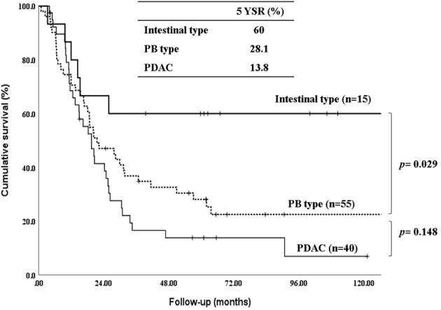 Figure 2
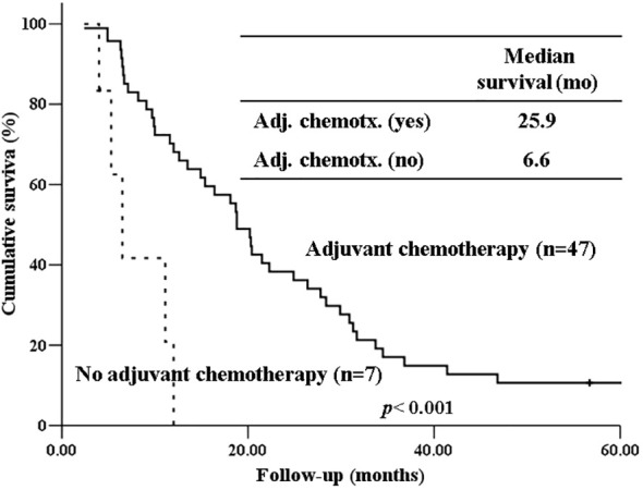 Figure 3
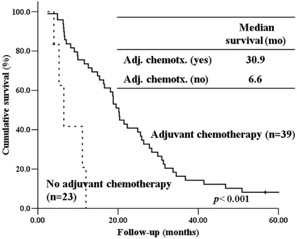 Figure 4
| Histologic Phenotype |
| ||
|---|---|---|---|
| Intestinal type (n=17) | Pancreatobiliary type (n=93) | ||
| Age, median, y | 64.0 ± 9.4 | 64.0 ± 9.1 | 0.984 |
| Gender (M:F) | 1.5:1 | 1.7:1 | 0.507 |
| Tumor origin | <0.001 | ||
| CBD | 8 (47.1%) | 39 (41.9%) | |
| Pancreas | 2 (11.7%) | 38 (41.2%) | |
| AoV | 6 (35.3%) | 11 (11.8%) | |
| Duodenum | 1 (5.9%) | 5 (5.4%) | |
| Tumor size (cm) | 3.7 ± 1.4 | 3.2 ± 1.7 | 0.665 |
| Elevation of preoperative serum CEA | 2 (14.3%) | 22 (27.8%) | 0.285 |
| Elevation of preoperative serum CA19-9 | 12 (70.6%) | 64 (68.8%) | 0.955 |
| Histologic grade | |||
| Well-differentiated | 6 (35.3%) | 12 (11.8%) | 0.001 |
| T stage | |||
| T1&2: T3&4 | 1:0.7 | 1:2.9 | 0.007 |
| T3&4 | 7 (41.2%) | 69 (74.2%) | 0.007 |
| Nodal status (N1&2) | 5 (29.4%) | 52 (55.9%) | 0.022 |
| PNI (Yes) | 7 (41.2%) | 76 (76.4%) | 0.029 |
| LVI (Yes) | 4 (23.5%) | 48 (36.4%) | 0.001 |
| Curative resection, R0 (Yes) | 15 (88.2%) | 78 (83.9%) | 0.514 |
| Adjuvant treatment (Yes) | 13 (76.5%) | 70 (75.3%) | 0.092 |
| Chemotherapy (Yes) | 10 (76.9%) | 39 (55.7%) | |
| Gemcitabine-based | 4 (40.0%) | 16 (41.0%) | |
| 5-FU-based | 2 (20.0%) | 18 (46.2%) | |
| Others | 4 (40.0%) | 5 (12.8%) | |
| Chemoradiotherapy (Yes) | 1 (7.7%) | 23 (32.9%) | |
| Radiotherapy (Yes) | 2 (15.4%) | 8 (11.4%) | |
| Recurrence (Yes) | 7 (41.2%) | 55 (59.1%) | 0.170 |
| Regional | 2 (28.6%) | 4 (7.1%) | 0.005 |
| Systemic | 5 (71.4%) | 51 (92.9%) | |
| Histologic Phenotype |
| |||
|---|---|---|---|---|
| Intestinal type | Pancreatobiliary | *PDAC | ||
| Age, median, y | 64.0 ± 9.4 | 64.0 ± 9.1 | 63.0 ± 8.3 | 0.984 |
| Gender (M:F) | 9:6 | 1:1 | 25:15 | 0.507 |
| Tumor origin | ||||
| CBD | 8 (17.0%) | 39 (83.0%) | <0.001 | |
| AoV | 6 (35.3%) | 11 (64.7%) | ||
| Duodenum | 1 (16.7%) | 5 (83.3%) | ||
| Tumor size (cm) | 3.7 ± 1.4 | 3.2 ± 1.7 | 3.1 ± 1.1 | 0.062 |
| Elevation of preoperative serum CEA | 2 (16.7%) | 9 (19.6%) | 13 (37.1%) | 0.149 |
| Elevation of preoperative serum CA19-9 | 10 (66.7%) | 32 (65.3%) | 32 (80.0%) | 0.288 |
| Histologic grade | ||||
| W/D | 5 (33.3%) | 8 (14.5%) | 4 (10.0%) | 0.005 |
| T stage | <0.001 | |||
| T1,2: T3,4 | 2:1 | 21:34 | 3:37 | |
| T3,4 | 5 (33.3%) | 34 (61.8%) | 37 (92.5%) | |
| Nodal status: N1,2 | 5 (33.3%) | 23 (41.8%) | 29 (72.5%) | 0.004 |
| PNI (Yes) | 7 (46.7%) | 42 (76.4%) | 34 (85.0%) | 0.029 |
| LVI (Yes) | 4 (26.7%) | 20 (36.4%) | 28 (70.0%) | 0.001 |
| Curative resection, R0 (Yes) | 13 (86.7%) | 43 (78.2%) | 35 (87.5%) | 0.450 |
| Adjuvant treatment (Yes) | 12 (80.0%) | 40 (72.7%) | 31 (77.5%) | 0.128 |
| Chemotherapy (Yes) | 9 (75.0%) | 21 (52.5%) | 19 (61.3%) | |
| Gemcitabine-based | 3 (33.3%) | 10 (47.6%) | 9 (47.4%) | |
| 5-FU-based | 2 (22.2%) | 7 (33.3%) | 9 (47.4%) | |
| Others | 4 (44.4%) | 4 (19.0%) | 1 (5.2%) | |
| Chemoradiotherapy (Yes) | 1 (8.3%) | 12 (30.0%) | 11 (35.5%) | |
| Radiotherapy (Yes) | 2 (16.6%) | 7 (17.5%) | 1 (3.2%) | |
| Recurrence (Yes) | 6 (40.0%) | 32 (58.2%) | 24 (60.0%) | 0.383 |
| Regional | 2 (33.3%) | 2 (6.3%) | 2 (8.0%) | 0.049 |
| Systemic | 4 (67.7%) | 30 (93.7%) | 22 (92.0%) | |
| variables | Univariate | Multivariate | ||||
|---|---|---|---|---|---|---|
| Subgroup | MST (mo) |
| HR | 95% CI |
| |
| Elevation of preoperative serum CEA | Yes | 21.2 | 0.024 | 1.286 | 0.585-2.828 | 0.897 |
| No | 60.0 | |||||
| Curative resection | R0 | 58.2 | 0.002 | 0.417 | 0.219-0.792 | 0.008 |
| R1,R2 | 16.8 | |||||
| Histologic type | I type | 105.8 | 0.003 | 0.402 | 0.231-1.801 | 0.333 |
| PB type | 41.8 | |||||
| Lymph node metastasis | Yes | 39.2 | 0.018 | 0.575 | 0.280-1.180 | 0.297 |
| No | 64.6 | |||||
| Perineural invasion | Yes | 37.7 | 0.003 | 0.545 | 0.197-1.508 | 0.519 |
| No | 97.5 | |||||
| Lymphovascular invasion | Yes | 38.9 | 0.004 | 0.854 | 0.425-1.715 | 0.978 |
| No | 63.8 | |||||
Peer Reviews
No public reviews on file for this paper yet. If you reviewed it on a platform where reviews are public (OpenReview, ICLR, NeurIPS, ICML), you can paste yours below so the community can read it here.
Videos
No videos yet. Explain this paper in a talk, walkthrough, or lecture? Add one.
Taxonomy
TopicsPancreatic and Hepatic Oncology Research · Cholangiocarcinoma and Gallbladder Cancer Studies · Genetic factors in colorectal cancer
Introduction
1
Periampullary cancer develops around the ampulla of Vater and originates anatomically from the pancreatic head, duodenum, distal bile duct, or ampulla. The World Health Organization (WHO) and American Joint Committee on Cancer (AJCC) have classified periampullary cancer into the above-mentioned four types according to the anatomical location and the tumor, node, metastasis (TNM) staging criteria are different for each origin. (1, 2), However, in the case of the duodenal, distal bile duct, and ampullary cancers, except for pancreatic head cancer which has the worst prognosis, it is often difficult to distinguish the exact site of origin owing to the destruction of normal periampullary anatomy or the presence of epithelial dysplasia even with standardized histopathologic evaluation. Furthermore, this makes it difficult to predict the prognosis and decide on adequate treatment, especially regimen of adjuvant chemotherapy, as the prognosis of patients after pancreaticoduodenectomy (PD) varies greatly even in the same anatomic location (3, 4),. Therefore, several recent studies have questioned this anatomical classification and addressed the need for a new classification (5–7).
Kimura et al. first reported dividing adenocarcinomas originating around the ampulla into intestinal and pancreaticobiliary (PB) types and demonstrated that the latter type had a worse prognosis (8). The intestinal type frequently resembles colon cancer and originates from the intestinal epithelium overlying the ampulla, evolving through an adenoma-dysplasia-adenocarcinoma sequence. In contrast, the PB type originates from the endothelium of the distal common bile duct and distal pancreatic intraepithelial neoplasia in an analogous dysplasia-adenocarcinoma sequence. Although the histologic phenotype in pancreatic ductal adenocarcinoma has not been endorsed by WHO until now, several groups have extended intestinal and pancreatobiliary concepts to the entire spectrum of periampullary adenocarcinoma including pancreatic ductal adenocarcinoma (9–13). In these studies, the intestinal type in pancreatic ductal adenocarcinoma accounted for approximately less than 10% (9–13). Histologic phenotype can be easily determined based on hematoxylin-eosin (H&E) staining. Several studies have shown that the histological type of periampullary adenocarcinomas has biological and prognostic relevance (9–15). However, some other studies have demonstrated no survival difference between the intestinal type and PB type, and the prognostic potential of the histological classification of periampullary cancer has been debated (16–18). These discrepancies between studies may be because of the low incidence of periampullary cancer excluding pancreatic cancer, and the consequent small cohorts with reliable data. Therefore, this study was designed to compare the clinicopathological characteristics and long-term outcomes between intestinal- and PB-type periampullary adenocarcinomas after curative-intent resection in a tertiary referral center to determine the potential of this histological classification as an independent prognostic factor.
Materials and methods
2
Study subjects
2.1
This study was approved by the Institutional Review Board of Chung-Ang University Hospital, Seoul, Korea. The requirement for written informed consent was waived because of the retrospective nature of the study. We retrospectively reviewed the medical records of patients who underwent curative-intended pancreatoduodenectomy for primary periampullary carcinoma from May 2010 to February 2020 at the Chung-Ang University Hospital, a tertiary referral hospital. Patients who underwent resection with macroscopic residual tumor (R2 resection) or a palliative procedure, and patients with distant metastasis and who diagnosed as other than adenocarcinoma (e.g., adenosquamous cell carcinoma, mucinous adenocarcinoma, and anaplastic type adenocarcinoma) were excluded. In addition, patients with fewer than 6 lymph nodes sampled were also excluded. Finally, a total of 110 patients were included in this study. All PDs were performed by senior pancreatic surgeons. Two expert pancreatic pathologists re-evaluated all pathological slides and determined their histological phenotypes.
Histological phenotype determination
2.2
The histological phenotype was determined as intestinal type or PB type based on the criteria first suggested by Kimura et al. and later revised by Saavedra et al. (8), (19), In brief, pancreatobiliary tumors typically feature simple or branching glands and small solid nests of cells surrounded by a desmoplastic stroma. They commonly exhibit cuboidal to low columnar epithelium arranged in a single layer without nuclear pseudostratification. The nuclei within these tumors are typically rounded, although there is notable variation in size and shape from one cell to the next. Intestinal tumors on the other hand, often resembling colon cancer, may present as solid nests with cribriform areas and are characterized by tall and often pseudostratified columnar epithelium. In these tumors, oval nuclei are typically located in the more basal aspects of the cytoplasm, and may also often contain mucin. Cases of mixed-type differentiation were classified according to the dominant pattern. In case of controversy regarding the histological phenotype, a consensus was reached by a second evaluation of the slides. Furthermore, all pathological slides were retrospectively reclassified according to the American Joint Committee on Cancer 8th edition definition.
The analyzed histopathological features included tumor size, histological grade, TNM stage, perineural invasion, and lymphovascular invasion. The tumor stage was defined according to the American Joint Committee on Cancer 8th edition definition. Perioperative medical records were reviewed for patient demographics, surgery type, follow-up, recurrence, and disease-specific survival. Overall survival was defined as the time from surgery to the last follow-up visit.
Statistical analysis
2.3
Continuous data are reported as means ± standard deviation (SDs), while numbers and percentages are used for categorical variables. Categorical variables were examined using the Chi-square test. Continuous variables such as maximum tumor diameter were compared using Mann-Whitney U test. Statistical significance was set at p < 0.05. The Kaplan–Meier method was used to calculate the curves for overall survival. Cox regression hazard models were used for multivariate analyses. The results are reported as hazard ratios (HR) with 95% confidence intervals (CI). A two-tailed p value <0.05 was considered statistically significant. All statistical analyses were performed using the SPSS version 25 for Windows (IBM Corp., Armonk, NY, USA).
Results
3
A total of 110 patients underwent curative-intended PD (R0 or R1 resection) and were pathologically diagnosed with periampullary adenocarcinoma at the Chung-Ang University Hospital between May 2010 and February 2020. The enrolled patients comprised 93 (84.5%) patients with PB type and 17 with intestinal-type differentiation. Of the total, 47 patients had extrahepatic bile duct adenocarcinoma, 40 had pancreatic adenocarcinoma, 17 had ampulla of Vater adenocarcinoma, and six had duodenal adenocarcinoma. The characteristics of each histological group are summarized in Table 1. The intestinal type had significantly more well-differentiated histology (35.3% vs. 11.8%, p=0.001), significantly less nodal metastasis (29.4% vs.55.9%, p=0.022), perineural invasion (41.2% vs. 76.4%, p=0.029), lymphovascular invasion (23.5% vs. 36.4%, p=0.001), and advanced T stage (more than T3; 41.2% vs.74.2%, p=0.007) than the PB type. There were no significant differences in the curative resection, adjuvant treatment rates, and recurrence rates. However, systemic recurrence (92.9% vs. 66.7%, p=0.005) was more common in the PB type than in the intestinal type.
Comparing intestinal type, PB types excluding pancreatic ductal adenocarcinoma (PDAC) and PDAC, PDAC group had significantly more advance T stage (more than T3; 33.3% vs. 61.8% vs. 92.5%, *p<*0.001), nodal metastasis (33.3% vs. 41.8% vs. 72.5%, *p=*0.004), perineural invasion (46.7% vs.76.4% vs.85.0%, *p=*0.029), lymphovascular invasion (26.7% vs.36.4% vs.70.0%, *p=*0.001) than the other two groups. (Table 2) There were no significant differences in curative resection rates, adjuvant chemotherapy and recurrence rates. For recurrence pattern, systemic recurrence was significantly more common in PB type and PDAC than intestinal type.
The 5-year-overall survival rate of the intestinal type was significantly better than that of the PB type (58.8% vs. 20.4%, *p=*0.003) (Figure 1), and all patients with intestinal-type ampulla of Vater cancer (n=6) survived for more than 5 years. The 5-year relapse-free survival rate of the intestinal type was also significantly better than that of the PB type (56.3% vs. 17.4%, *p=*0.004). There was no significant difference in the 5-year-overall survival rate between the PB types excluding PDAC and PDAC (39.4% vs. 19.2%, *p=*0.148) (Figure 2). There was also no significant difference in the 5-year relapse-free survival rate between the PB types excluding PDAC and PDAC (22.3% vs. 10.9%, *p=*0.289). For patients with systemic recurrence, adjuvant chemotherapy significantly improved survival (median survival, 25.9 mo vs. 6.6 mo, p<0.001) (Figure 3). However, there was no significant difference between chemotherapy regimens. Furthermore, for patients with PB type with recurrence, adjuvant chemotherapy significantly improved patient survival (median survival, 30.9 mo vs. 6.6 mo, p<0.001) (Figure 4).
Kaplan-Meier survival curves comparing the overall survival of patients after resection of periampullary adenocarcinomas grouped by histopathologic phenotype.
Kaplan-Meier survival curves comparing the overall survival of patients after resection of periampullary adenocarcinomas grouped by Intestinal type, PB types excluding pancreatic ductal adenocarcinoma and pancreatic ductal adenocarcinoma.
Kaplan-Meier survival curves comparing the overall survival of patients who experienced systemic recurrence depending on whether receiving adjuvant chemotherapy or not.
Kaplan-Meier survival curves comparing the overall survival of patients with PB type histology who experienced recurrence depending on whether receiving adjuvant chemotherapy or not.
In the univariate analysis, the intestinal type showed a longer mean survival time (MST) than that of the PB type (105.8 months vs. 41.8 months). Non-curative resection (16.8 months vs. 58.2 months, *p=*0.002), presence of lymph node metastasis (39.2 months vs. 64.6 months, *p=*0.018), perineural invasion (37.7 months vs. 97.5 months, *p=*0.003), and lymphovascular invasion (38.9 months vs. 63.8 months, *p=*0.004) showed shorter MST. However, in the multivariate analysis, only curative resection was significantly associated with survival (hazard ratio, 0.417; 95% CI, 0.219-0.792, p=0.008). Other factors, such as tumor location, histologic subtype, T stage, N stage, and lymph node metastasis were not significant. (Table 3).
Discussion
4
Despite the same anatomical location, periampullary cancers show variable prognoses and responses to adjuvant therapy after tumor resection. Histologic subtyping of periampullary cancers into intestinal and PB types is emerging as an alternative to the current classification of anatomic location. This is because histologic subtyping is thought to have the potential to predict the biological characteristics and prognosis of cancer better than the conventional classification based on the anatomical origin of the tumor. Furthermore, some studies have shown that adjuvant systemic therapy improves overall survival in patients with the PB type, mimicking pancreatic ductal adenocarcinoma, whereas the intestinal type does not benefit from the same regimen (20, 21). Therefore, we hypothesized that histopathologic tumor characteristics and long-term survival after curative–intended pancreatoduodenectomies for patients with PB type cancer might be similar to pancreatic ductal adenocarcinoma and have a worse prognosis than the intestinal type.
In the present study, we found that intestinal-type cancers had lower pathological aggressiveness and better prognoses than the PB type. In contrast, the PB type showed clinicopathological features and prognosis similar to those of pancreatic ductal adenocarcinoma. The PB type had significantly more aggressive histology, nodal metastasis, perineural invasion, lymphovascular invasion, advanced T stage, and systemic metastasis than the intestinal type while the PB type and pancreatic ductal adenocarcinoma showed similar aggressive pathological features and poor overall survival. These results further strengthen the consistency of clinicopathological features of histological phenotypes and the possibility of histological classification as a prognostic factor for periampullary cancers rather than anatomic location. A meta-analysis of 37 studies on histological subtype demonstrated similar results to our study (22). PB type had significantly higher rates of advanced T stage, lymph node metastasis, poor tumor differentiation, lymphovascular invasion, perineural invasion, and worse survival than intestinal type. Although most of the studies included in this meta-analysis were focused on ampullary cancer, recently, intestinal type, not only in ampullary cancers but also in pancreatic ductal adenocarcinoma, has shown significantly better survival than PB type (9–12). This had not been demonstrated before due to the relatively rare occurrence of intestinal type in pancreatic ductal adenocarcinoma. Specifically, Westgaard, et al. demonstrated that long-term survival curves of intestinal type of four periampullary tumors, including pancreatic ductal adenocarcinoma, were very similar regardless of tumor location when adjusted for tumor diameter and nodal status (11). Therefore, in cases of periampullary cancer, we suggest determining the histological subtype routinely, in addition to the classification of anatomical location. In most cases, the histological phenotype can be determined by hematoxylin and eosin (H&E) staining alone and may provide more information about adjuvant treatment and prognosis. For morphologically challenging cases, addition of small battery of immunohistochemical markers can improve accuracy (15, 23). A recently published meta-analysis also recommended routine histopathological subtype classification, even in a clinical setting without immunohistochemistry, because the histopathological subtype is a major prognostic factor (22).
Our study also showed that systemic metastasis were more common in the PB type than in the intestinal type. For patients with recurrence, especially those with systemic recurrence, adjuvant chemotherapy significantly improved survival (median survival, 25.9 mo vs. 6.6 mo, p<0.001). Furthermore, for patients with PB type with recurrence, adjuvant chemotherapy significantly improved patient survival (median survival, 30.9 mo vs. 6.6 mo, p<0.001). Therefore, adjuvant chemotherapy should be considered more actively for PB type cancer. Due to the low number of patients per chemotherapeutic regimen in our study, we were unable to determine if there are differences in patient survival based on specific chemotherapeutic regimens. To date, only a few randomized clinical trials have been performed to demonstrate the role of adjuvant chemotherapy after the resection of periampullary adenocarcinoma, excluding pancreatic ductal adenocarcinoma, because the incidence of each tumor is still low (24, 25).. However, several recent studies have shown that gemcitabine-based adjuvant chemotherapy is associated with prolonged overall survival in the PB type of ampullary cancer but not in the intestinal type (26, 27). There has been no study yet comparing the effects of adjuvant chemotherapy between intestinal type and PB type in pancreatic ductal adenocarcinoma. Future studies should evaluate the validity of stratification by histologic type of periampullary cancers in adjuvant treatment. Therefore, when selecting an adjuvant chemotherapy regimen, the histological subtype should be considered.
An additional issue with histologic type of these cancers is effectiveness of metastatectomy in intestinal type. Given the intestinal type is associated with less aggressive histolopathologic feature, a prior study showed that resection of periampullary adenocarcinoma liver metastasis provides survival benefit only in intestinal type. Considering the benefit of hepatic metastatectomy for the colon cancer and similarity of intestinal type to colon cancer, further study on metastatectomy in intestinal type should be performed near future.
This study had some limitations. First, it was a retrospective, single-center study, leading to a possible selection bias. Second, the sample size may have been too small and unbalance of sample size between PB type and intestinal type may have been too big to show statistical significance or draw definitive conclusions. However, because periampullary cancers are uncommon malignancies and prospective studies are difficult to conduct, a well-organized retrospective study would be a reasonable alternative.
Conclusions
5
Although on multivariate analysis, histologic subtype was not an independent prognostic factor because of the small number of included patients, histologic phenotypes, including intestinal and PB types, showed the possibility of being prognostic factors in periampullary cancer. Because of the profound differences in clinical biology and prognosis, the histological classification of periampullary cancer into PB and intestinal types should be encouraged to predict prognosis and choose optimal adjuvant treatment. Further large-scale studies are required to demonstrate the prognostic significance of the histologic phenotype.
Data availability statement
The data presented in this study are available on request from the corresponding author. The data are not publicly available due to database complexity and local storage guideline.
Ethics statement
The studies involving humans were approved by Chung-Ang University Hospital Institutional Review Board (IRB No. 1961-003-365). The studies were conducted in accordance with the local legislation and institutional requirements. The ethics committee/institutional review board waived the requirement of written informed consent for participation from the participants or the participants' legal guardians/next of kin because of the retrospective nature of the study and the patients have already passed.
Author contributions
H-SK: Conceptualization, Data curation, Formal Analysis, Writing – original draft. C-MH: Conceptualization, Data curation, Formal Analysis, Investigation, Methodology, Writing – original draft. Y-SC: Methodology, Software, Validation, Writing – review & editing. S-WS: Methodology, Resources, Software, Visualization, Writing – review & editing. SL: Conceptualization, Formal Analysis, Project administration, Supervision, Validation, Visualization, Writing – original draft, Writing – review & editing.
The reference list from the paper itself. Each links out to its DOI / PubMed record.
- 1Bosman FT Carneiro F Hruban RH Theise ND. WHO Classification of Tumors of the Digestive System. 4th ed. Lyon, France: International Agency for Research in Cancer (2010).
- 2Edge SB Byrd DR Compton CC Fritz AG Greene FL Trotti A. AJCC Cancer Staging Manual. 7th ed. New York, NY: Springer (2010).
- 3Frierson HF Jr. The gross anatomy and histology of the gallbladder, extrahepatic bile ducts, Vaterian system, and minor papilla. Am J Surg Pathol. (1989) 13:146–62. doi: 10.1097/00000478-198902000-00008 2644853 · doi ↗ · pubmed ↗
- 4Van Geenen RC Van Gulik TM Offerhaus GJ De Wit LT Busch OR Obertop H. Survival after pancreaticoduodenectomy for periampullary adenocarcinoma: an update. Eur J Surg Oncol. (2001) 27:549–57. doi: 10.1053/ejso.2001.1162 11520088 · doi ↗ · pubmed ↗
- 5Adsay NV Bagci P Tajiri T Oliva I Ohike N Balci S. Pathologic staging of pancreatic, ampullary, biliary, and gallbladder cancers: pitfalls and practical limitations of the current AJCC/UICC TNM staging system and opportunities for improvement. Semin Diagn Pathol. (2012) 29:127–41. doi: 10.1053/j.semdp.2012.08.010 23062420 · doi ↗ · pubmed ↗
- 6Adsay V Ohike N Tajiri T Kim GE Krasinskas A Balci S. Ampullary region carcinomas: definition and site specific classification with delineation of four clinicopathologically and prognostically distinct subsets in an analysis of 249 cases. Am J Surg Pathol. (2012) 36:1592–608. doi: 10.1097/PAS.0b 013e 31826399 d 8 23026934 · doi ↗ · pubmed ↗
- 7Williams JL Chan CK Toste PA Elliott IA Vasquez CR Sunjaya DB. Association of histopathologic phenotype of periampullary adenocarcinomas with survival. JAMA Surg. (2017) 152:82–8. doi: 10.1001/jamasurg.2016.3466 27732711 · doi ↗ · pubmed ↗
- 8Kimura W Futakawa N Yamagata S Wada Y Kuroda A Muto T. Different clinicopathologic findings in two histologic types of carcinoma of papilla of Vater. Jpn J Cancer Res. (1994) 85:161–6. doi: 10.1111/j.1349-7006.1994.tb 02077.x PMC 59194257511574 · doi ↗ · pubmed ↗