The characteristic “alveolus nest sign” in solid pseudopapillary neoplasm of the pancreas
Weigang Gu, Justin Ryan L. Tan, Hang Bin Jin, Quifeng Lou, Ka Shing Cheung, Jianfeng Yang, Xiaofeng Zhang

Abstract
Genes, proteins, chemicals, diseases, species, mutations and cell lines named across the full text — each resolved to its canonical identifier and authoritative record.
Click any figure to enlarge with its caption.
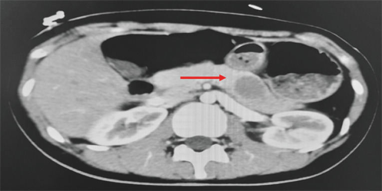 Fig. 1
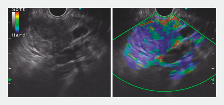 Fig. 2
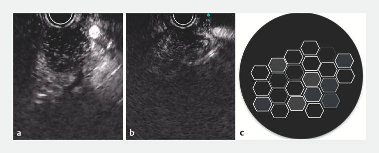 Fig. 3
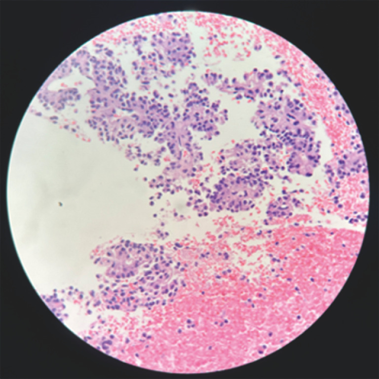 Fig. 4
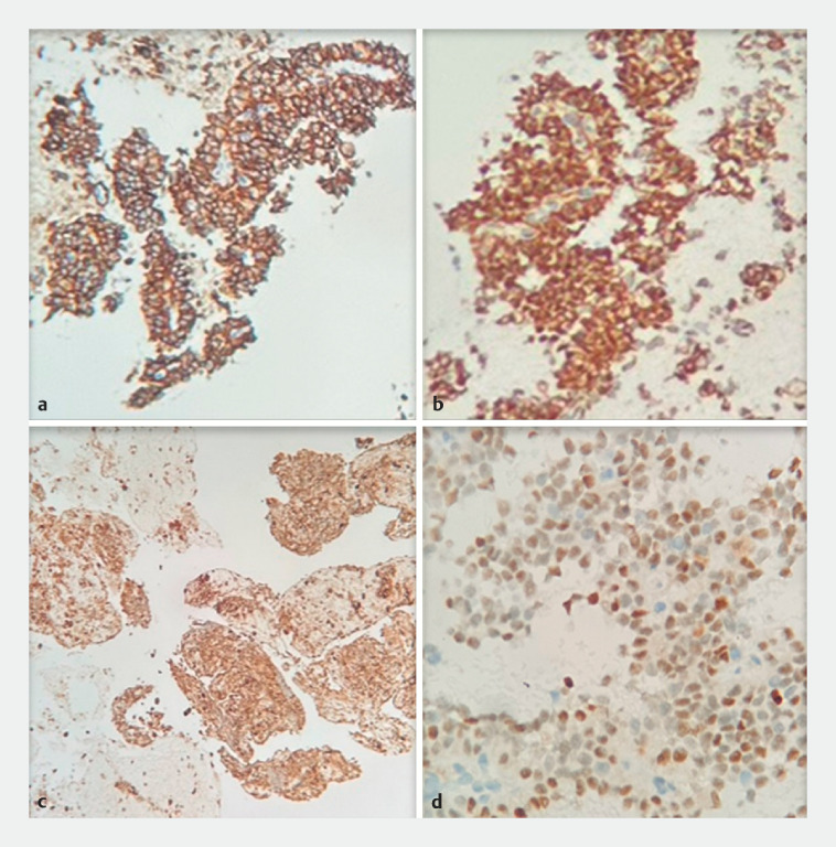 Fig. 5
- —The Natural Science Foundation of Zhejiang Province
- —Zhejiang Medical and Health Science and Technology Plan
- —Zhejiang Province’s 2023 Key R&D Plan Project
- —Hangzhou Medical and Health Science and Technology Plan
Peer Reviews
No public reviews on file for this paper yet. If you reviewed it on a platform where reviews are public (OpenReview, ICLR, NeurIPS, ICML), you can paste yours below so the community can read it here.
Videos
No videos yet. Explain this paper in a talk, walkthrough, or lecture? Add one.
Taxonomy
TopicsPancreatic and Hepatic Oncology Research · Pancreatitis Pathology and Treatment · Gastrointestinal disorders and treatments
A 23-year-old woman presented with a 1-week history of abdominal pain radiating to the back. The pain was not relieved by proton pump inhibitors. Contrast-enhanced abdominal computed tomography scan demonstrated a round, well-defined, hypodense lesion within the pancreatic body ( Fig. 1 ). On endoscopic ultrasound (EUS) elastography there was a 2 × 2.5-cm hypoechoic mass that was predominantly hard (blue) with dispersed heterogeneous soft (green) areas ( Fig. 2 ). Contrast-enhanced harmonic endoscopic ultrasound (CH-EUS) revealed hyperechoic solid granular components interspersed with small anechoic regions within the pancreatic mass ( Fig. 3 ). These characteristics were typical of the “alveolus nest sign” and were present during the arterial and venous phases of CH-EUS ( Video 1 ). EUS-guided fine-needle biopsy with subsequent histopathologic examination ( Fig. 4 ) and immunohistochemical analysis ( Fig. 5 ) yielded a definitive diagnosis of solid pseudopapillary neoplasm (SPN). The patient underwent distal pancreatectomy for definitive treatment.
Contrast-enhanced computed tomography scan revealed a round, well-defined, hypodense lesion (red arrow) within the pancreatic body.
Endoscopic ultrasound elastography revealed a 2 × 2.5-cm hypoechoic mass that was predominantly hard (blue) with dispersed heterogeneous soft (green) areas.
Contrast-enhanced harmonic endoscopic ultrasound revealed hyperechoic solid granular components interspersed with small anechoic regions within the pancreatic mass. a Arterial phase. b Venous phase. c Visual representation of the alveolus nest sign.
Histopathology specimen showed scattered epithelioid cells with clear or eosinophilic cytoplasm arranged in sheets or papillary shapes.
Immunohistochemical tests showed the following positive results. a CD56. b β-catenin. c α-1-antitrypsin. d Progesterone receptor.
The alveolus nest sign during the arterial and venous phases of contrast-enhanced harmonic endoscopic ultrasound.Video 1
SPN is an uncommon pancreatic neoplasm exhibiting low malignant potential. It predominantly affects young females and is characterized by indolent behavior, with an excellent long-term prognosis for the majority of patients 1 2 3 4 . CH-EUS examination may reveal characteristic features suggestive of SPN pathology, including the presence of an intralesional pseudopapillary architecture or intratumoral clefting, also termed the “alveolus nest sign” 1 2 . On CH-EUS, the alveolus nest sign is seen as a region with isoechogenicity to hyperechogenicity, containing a solid and granular component. This component is interspersed with multiple small anechoic areas of varying sizes. These anechoic regions become evident on CH-EUS typically 40 seconds after contrast injection. The presence of this sign at any time within 40 seconds to 5 minutes post-contrast administration is considered characteristic of the alveolus nest sign 1 2 . CH-EUS is useful in differentiating SPN of the pancreas from other pancreatic tumors. Alveolus nest sign is a characteristic feature of SPN on CH-EUS.
Endoscopy_UCTN_Code_CCL_1AF_2AZ
The reference list from the paper itself. Each links out to its DOI / PubMed record.
- 1Ishikawa T Itoh A Kawashima HA case of solid-pseudopapillary neoplasm, focusing on contrast-enhanced endoscopic ultrasonography J Med Ultrason (2001)20113820921610.1007/s 10396-011-0313-z 27278586 · doi ↗ · pubmed ↗
- 2Kataoka K Ishikawa T Ohno E Differentiation between solid pseudopapillary neoplasm of the pancreas and nonfunctional pancreatic neuroendocrine neoplasm using endoscopic ultrasound Pancreas 20225110611135195603 10.1097/MPA.0000000000001966 · doi ↗ · pubmed ↗
- 3Papavramidis T Papavramidis S Solid pseudopapillary tumors of the pancreas: review of 718 patients reported in English literature J Am Coll Surg 200520096597210.1016/j.jamcollsurg.2005.02.01115922212 · doi ↗ · pubmed ↗
- 4Kim MJ Choi DW Choi SH Surgical treatment of solid pseudopapillary neoplasms of the pancreas and risk factors for malignancy Br J Surg 20141011266127110.1002/bjs.957725052300 · doi ↗ · pubmed ↗