Mechanical failure of a curved linear array echoendoscope: loose fixation of the high frequency transducer
Junichi Kaneko, Hiroki Tamakoshi, Tomoyuki Niwa, Masaki Takinami, Masafumi Nishino, Yurimi Takahashi, Takanori Yamada

Abstract
Genes, proteins, chemicals, diseases, species, mutations and cell lines named across the full text — each resolved to its canonical identifier and authoritative record.
Click any figure to enlarge with its caption.
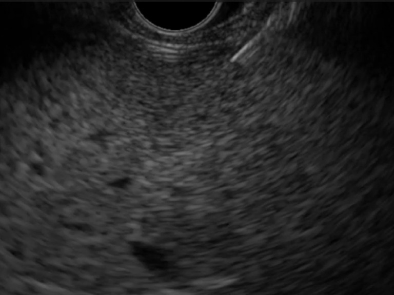 Fig. 1
 Fig. 2
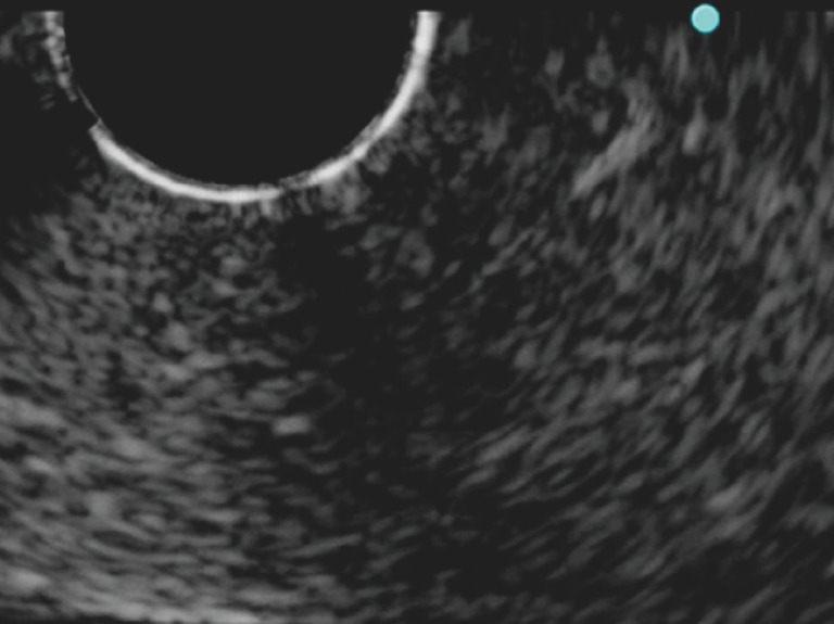 Fig. 3
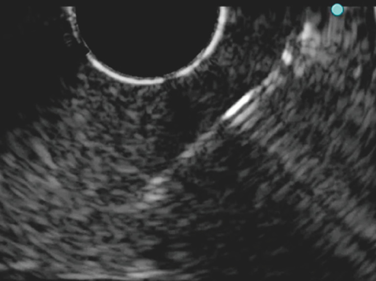 Fig. 4
Peer Reviews
No public reviews on file for this paper yet. If you reviewed it on a platform where reviews are public (OpenReview, ICLR, NeurIPS, ICML), you can paste yours below so the community can read it here.
Videos
No videos yet. Explain this paper in a talk, walkthrough, or lecture? Add one.
Taxonomy
TopicsEsophageal and GI Pathology · Pancreatic and Hepatic Oncology Research · Gallbladder and Bile Duct Disorders
Endoscopic ultrasound (EUS), a well-established diagnostic modality, allows for visualization of previously inaccessible anatomical regions and enables tissue acquisition and therapeutic procedures 1 2 . In interventional EUS, a curved linear array echoendoscope is first used to visualize an object with a high frequency transducer at the echoendoscope tip. A dedicated needle is then used to puncture the object under ultrasound guidance 3 . If fixation of the high frequency transducer becomes loose and rotates, it becomes difficult to visualize the puncture needle. We describe a case and in vitro studies showing the effect of mechanical failure of an echoendoscope ( Video 1 ).
A case and in vitro studies showing the effects of mechanical failure of the echoendoscope in which fixation of the high frequency transducer is loose and rotated 10° counterclockwise.Video 1
A 76-year-old man was referred to our hospital with elevated liver enzyme levels, so an EUS-guided liver biopsy was planned. A curved linear array echoendoscope (GF-UCT260; Olympus Medical Systems Corp., Tokyo, Japan) and 19-gauge Franseen needle (Acquire; Boston Scientific Corporation, Natick, Massachusetts, USA) were used. First, the left liver lobe was visualized under ultrasonographic guidance. The sheath was recognized; however, the needle was obscured ( Fig. 1 ). During puncture, the needle was visualized by moving the endoscope. Finally, liver tissue was acquired without complications. After the procedure, fixation of the high frequency transducer was found to be loose and rotated 10° counterclockwise ( Fig. 2 ). Subsequent puncture experiments were performed using tofu. The puncture needle was poorly visualized when the high frequency transducer was rotated, but it was well visualized when the rotation was restored to normal ( Fig. 3 , Fig. 4 ).
The puncture needle is not visible during endoscopic ultrasound-guided liver tissue acquisition. Subsequently, it was found that fixation of the high frequency transducer was loose and rotated 10° counterclockwise.
An echoendoscope with the high frequency transducer rotated 10° counterclockwise.
Ultrasound image showing that visibility of the puncture needle was poor when the high frequency transducer was rotated 10° counterclockwise.
Ultrasound image showing that visibility of the puncture needle improved after correcting the position of the high frequency transducer.
The high frequency transducer was firmly fixed when this echoendoscope was purchased, and there were no instances of strong external shock waves. The cause of mechanical failure is unknown but may be due to age-related deterioration. In interventional EUS, loose fixation not only leads to an unsuccessful procedure but can also cause serious complications. Endoscopists should ensure proper fixation of the high frequency transducer before performing interventional EUS.
Endoscopy_UCTN_Code_TTT_1AS_2AF
The reference list from the paper itself. Each links out to its DOI / PubMed record.
- 1Kitano M Yoshida T Itonaga M Impact of endoscopic ultrasonography on diagnosis of pancreatic cancer J Gastroenterol 201954193210.1007/s 00535-018-1519-230406288 PMC 6314985 · doi ↗ · pubmed ↗
- 2Bojan K Peter VEUS tissue acquisition: From A to B Endosc Ultrasound 2020922523132655082 10.4103/eus.eus_21_20PMC 7528999 · doi ↗ · pubmed ↗
- 3Van Der Merwe SW Van Wanrooij RLJ Bronswijk M Therapeutic endoscopic ultrasound: European Society of Gastrointestinal Endoscopy (ESGE) Guideline Endoscopy 20225418520510.1055/a-1717-139134937098 · doi ↗ · pubmed ↗