Epithelioid Hemangioma Masquerading as a Clavicular Mass: A Diagnostic Conundrum
Pawani P Duddu, Inuganti Venkata Renuka, Santhi Imandi, Haritha Shah

TL;DR
A rare benign tumor called epithelioid hemangioma was diagnosed in a 40-year-old man after he presented with a clavicular mass.
Contribution
This case adds to the limited medical literature on epithelioid hemangioma and highlights its diagnostic challenges.
Findings
Epithelioid hemangioma was confirmed via histopathological examination of an excised clavicular mass.
The tumor was located at the medial end of the right clavicle and measured 3 x 2.5 cm.
Abstract
Epithelioid hemangioma (EH), a rare benign tumor, is documented in only a few cases in the medical literature. The complications of epithelioid hemangioma depend on the site of their presentation. If located in the eye, epithelioid hemangiomas may lead to amblyopia; in the larynx, they can cause respiratory issues; and if affecting bone, they can result in osteolytic lesions. Here, we present a case of a 40-year-old male who presented with swelling in the medial end of the right clavicle. Following clinical evaluation and a computed tomography scan, an excision biopsy was performed. The excised lesion measured 3 x 2.5 cm, and histopathological examination confirmed the finding as an epithelioid hemangioma.
Genes, proteins, chemicals, diseases, species, mutations and cell lines named across the full text — each resolved to its canonical identifier and authoritative record.
Click any figure to enlarge with its caption.
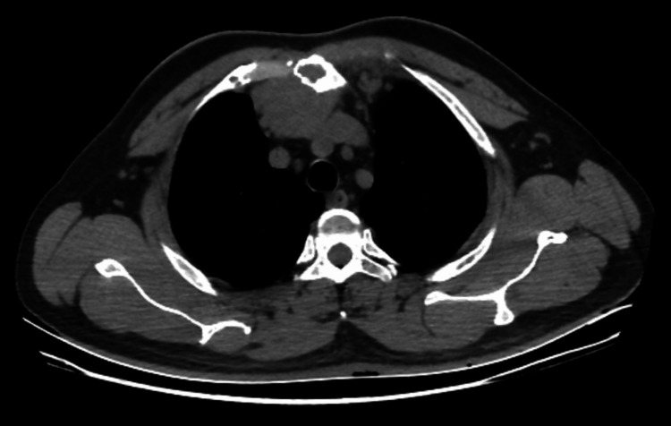 Figure 1
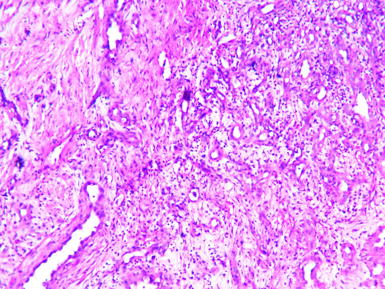 Figure 2
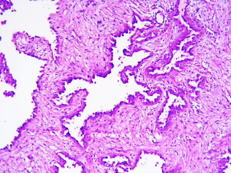 Figure 3
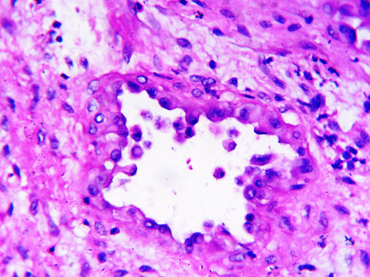 Figure 4
| No. | Study | Age (years)/Sex | Size (cm) | Site | Clinical Presentation | Imaging Findings | Histopathology | Treatment |
| 1 | Ángeles et al. [ | 32/M | 1.3x0.8 | Humeral artery | Lump on the right mid-arm | MRI: nodule formation is seen close to humeral artery and vein; USG: hyperechoic nodular lesion having smooth edges in contact with brachial artery | The tumor shows high vascularity and is infiltrated by epithelioid cells | Bypass with basilic branch graft |
| 2 | Nangia et al. [ | 30/F | 2x1 | Lingual alveolar mucosa | Painful, progressive swelling in the lingual gum tissue | Radiographs: the alveolar bone remained undamaged and unaffected by the mass positioned above it | The connective tissue encircled by epithelioid cells alongside a dense presence of inflammatory cells around the vessels | Excision of lesion |
| 3 | Wiggins et al. [ | 16/M | 15x5 | Scapular region | Painless, growing mass over left scapula | USG: highly vascularised intramuscular mass; MRI: intramuscular mass, prominent blood vessels observed | Spindle-shaped cells and robust endothelial cells | Excision, tissue rearrangement |
| 4 | Present Case | 40/M | 3x2.5 | Right clavicle | Painful swelling in the medial end of right clavicle | CECT chest: a well-defined heterogeneously enhanced mass at manubrium sternum | Lobular tumor composed of central epithelioid cells, and blood vessels lined by large oval cells with abundant eosinophilic cytoplasm and vesicular nuclei | Excision biopsy |
Peer Reviews
No public reviews on file for this paper yet. If you reviewed it on a platform where reviews are public (OpenReview, ICLR, NeurIPS, ICML), you can paste yours below so the community can read it here.
Videos
No videos yet. Explain this paper in a talk, walkthrough, or lecture? Add one.
Taxonomy
TopicsVascular Tumors and Angiosarcomas · Ocular Oncology and Treatments · Histiocytic Disorders and Treatments
Introduction
Hemangiomas are rare benign vascular neoplasms most commonly found in the head and neck, primarily on the forehead, as well as in the oral cavity, bones, and lymph nodes [1]. They are characterized by blood vessels surrounded by eosinophilic, epithelioid endothelial cells, and various inflammatory cells [1]. Trauma to bones or blood vessels may trigger their development, suggesting a potential underlying pathology [2]. Although hemangiomas are rare and lack a strong predisposition, adults show a slight tendency to develop this condition [3]. The clinical presentation varies depending on the site and may include subcutaneous nodules, abdominal discomfort, bone fractures, or even respiratory difficulties [3]. In this case report, we outline the clinical progression, diagnosis, and treatment of a case of epithelioid hemangioma in a patient who presented with a swelling in the medial end of the right clavicle.
Case presentation
A 40-year-old Indian male presented to the outpatient department with a painful swelling at the medial end of the right clavicle for one month. The patient had no record of any co-morbidities, such as diabetes or hypertension, and no history of any past surgeries. The patient was not on any medication prior to his visit to the hospital.
During clinical examination, a tender, palpable mass was identified over the medial end of the right clavicle. Laboratory tests showed leukocytosis with peripheral eosinophilia, while all other laboratory results were within normal ranges. A contrast-enhanced computed tomography (CECT) of the chest showed a well-defined, heterogeneously enhanced soft tissue mass measuring 4.6 x 4.4 x 4 cm on the right side of the manubrium sternum (Figure 1). It extended cranio-caudally from the D1 to D4 vertebral body. A computed tomography (CT)-guided fine needle aspiration cytology (FNAC) was performed to confirm the findings observed in the CECT scan and support the diagnosis of epithelioid hemangioma.
Contrast-enhanced computed tomography (CECT) of the chestA well-defined, heterogeneously enhanced soft tissue mass on the right side of the manubrium sterni.
An excisional biopsy was performed on the lesion and sent for histopathological examination. On examination, a nodular, gray-brown lesion measuring 3 x 2.5 cm was identified, showing gray-white areas on the cut-section. Microscopically, minimal to no mitotic activity was observed, although a hypocellular and fibrous stroma was present (Figure 2). Additionally, scant connective tissue was observed in the stroma (Figure 3), and blood vessels were lined by plump cells with vesicular nuclei and abundant eosinophilic cytoplasm (Figure 4).
Microphotographs of epithelioid hemangiomaShowing blood vessels and fibrous stroma (H&E scanner view).
Microphotographs of epithelioid hemangiomaBlood vessels lined by plump endothelial cells show scant fibrous tissue stroma (H&E x 100).
Microphotographs of epithelioid hemangiomaShowing blood vessels lined by plump cells with vesicular nuclei and abundant eosinophilic cytoplasm (H&E x 400).
Discussion
Epithelioid hemangioma was initially described as angiolymphoid hyperplasia with eosinophilia [1]. It can manifest in various sites, such as the eye, larynx, or bone [1]. The latter site was observed in our case. Based on recurrence and lymph node involvement, it can be classified as a more malignant or benign condition [4]. The tumor described in our case was benign in nature. Epithelioid hemangioma is characterized by blood vessels surrounded by plump, eosinophilic endothelial cells [1]. In our case, we observed blood vessels lined by plump cells with vesicular nuclei and abundant eosinophilic cytoplasm. These lesions may be localized to a single system or present at multiple sites simultaneously. In our case, there was a solitary lesion confined to the bone of the right clavicle.
Diagnosis involves a combination of clinical correlation, imaging, and biopsy, with biopsy considered the gold standard method [3]. In our case, clinical examination, coupled with contrast-enhanced computed tomography, revealed a well-defined, heterogeneously enhanced mass at the manubrium sternum (Table 1). These findings, combined with the biopsy report, correlated with the observations and led to the diagnosis of epithelioid hemangioma.
The treatment approach for epithelioid hemangioma varies depending on the individual case. In some asymptomatic cases, simple monitoring of the lesion may be sufficient, as it can regress over time [4]. However, in symptomatic cases, procedures such as curettage, embolization, or excisional biopsy may be performed to mitigate potential complications. In our case, we opted for an excisional biopsy of the lesion. The patient did not report any postoperative complaints.
Two years later, the patient returned with similar complaints, and a histopathological examination confirmed a recurrence of epithelioid hemangioma without any features of malignancy. However, the patient was lost to follow up.
Conclusions
We report a rare case of epithelioid hemangioma of bone in a patient who presented with a mass and associated tenderness at the medial end of the right clavicle. We opted for excisional biopsy as the treatment of choice, and the patient experienced no postoperative complications. However, the patient had a recurrence of epithelioid hemangioma with no features of malignancy two years later. The patient was subsequently lost to follow-up.
The reference list from the paper itself. Each links out to its DOI / PubMed record.
- 1Multiple epithelioid hemangiomas with orbital involvement Case Rep Pathol MiličićB Velnar T Pregelj R Limbaeck-Stokin C 629805201520152580278710.1155/2015/629805 PMC 4329744 · doi ↗ · pubmed ↗
- 2Epithelioid hemangioma of bone and soft tissue: a reappraisal of a controversial entity Clin Orthop Relat Res Errani C Zhang L Panicek DM Healey JH Antonescu CR 1498150647020122194830910.1007/s 11999-011-2070-0PMC 3314752 · doi ↗ · pubmed ↗
- 3Epithelioid hemangioma of bone: a rare vascular neoplasm. A case report and literature review J Long Term Eff Med Implants Savvidou O Korkolopoulou P Lakiotaki E 47553220223601792710.1615/J Long Term Eff Med Implants.2022041868 · doi ↗ · pubmed ↗
- 4Epithelioid haemangioma of bone: a case series and comprehensive literature review reappraising the diagnostic classification of all epithelioid vascular neoplasms of bone Cureus Ramkumar S 013202110.7759/cureus.15371 PMC 816862934094789 · doi ↗ · pubmed ↗
- 5Epithelioid hemangioma of the humeral artery: case report Annals of Clinical Case Reports Ángeles AR del Rosario MOM Enrique GT Carmen Nieves HL Daniel BM 214672022
- 6Epithelioid hemangioma of lingual alveolar mucosa: an immunohistochemical case report Case Rep Med Nangia R Puri A Gupta R Bansal S Negi A Chauhan I 436240201420142471591010.1155/2014/436240 PMC 3970468 · doi ↗ · pubmed ↗
- 7Atypical presentation and management of an epithelioid hemangioma: a case report and review of the literature Annals of Pediatric Surgery Wiggins CJ Dibbs RP Bartlett EL Ashton DJ Maricevish RS 53162020