Technical tips for antegrade endopancreatic radiofrequency ablation for severe pancreatojejunal stricture
Takeshi Ogura, Kimi Bessho, Nobuhiro Hattori, Jun Matsuno, Hiroki Nishikawa

Abstract
Genes, proteins, chemicals, diseases, species, mutations and cell lines named across the full text — each resolved to its canonical identifier and authoritative record.
Click any figure to enlarge with its caption.
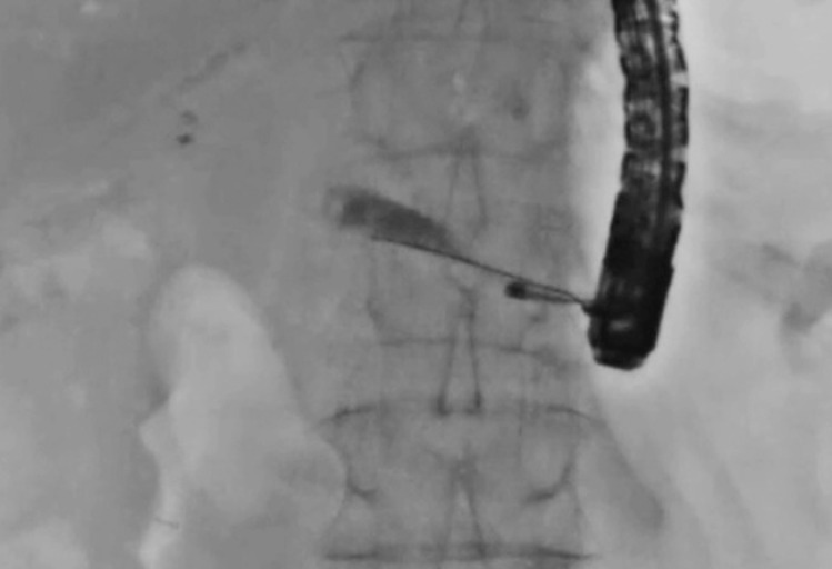 Fig. 1
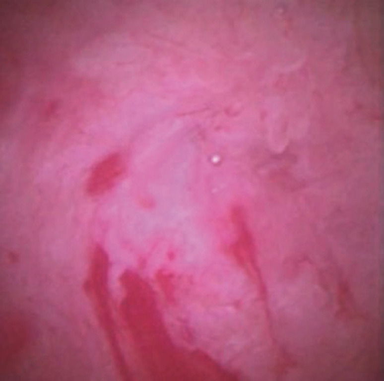 Fig. 2
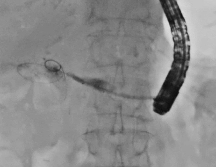 Fig. 3
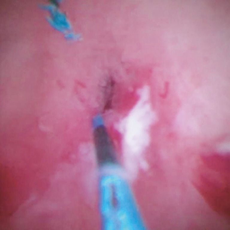 Fig. 4
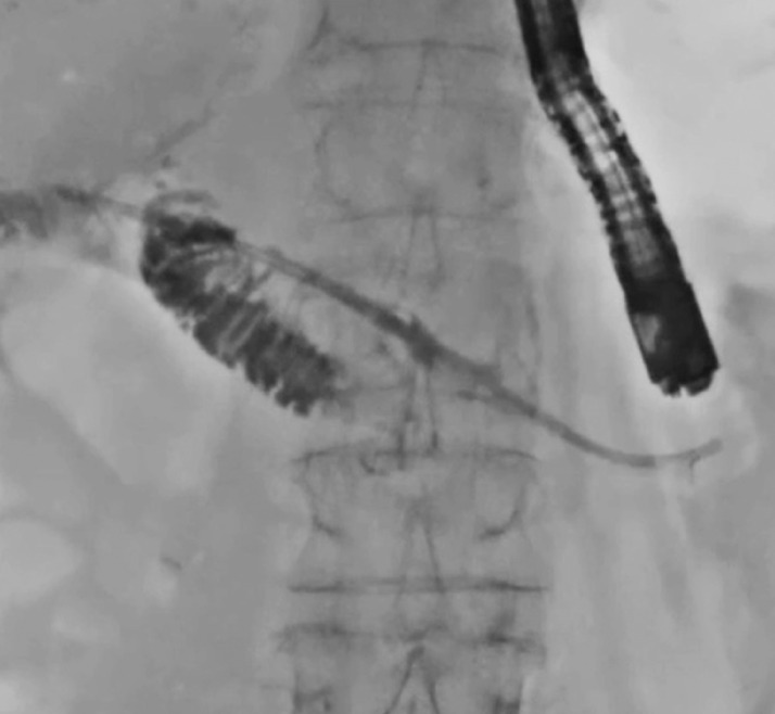 Fig. 5
Peer Reviews
No public reviews on file for this paper yet. If you reviewed it on a platform where reviews are public (OpenReview, ICLR, NeurIPS, ICML), you can paste yours below so the community can read it here.
Videos
No videos yet. Explain this paper in a talk, walkthrough, or lecture? Add one.
Taxonomy
TopicsPancreatic and Hepatic Oncology Research · Gallbladder and Bile Duct Disorders · Pancreatitis Pathology and Treatment
Pancreatojejunal stricture (PJS) is one of the late adverse events after pancreatoduodenectomy, and can lead to pancreatitis or endocrine pancreatic insufficiency as complications 1 . PJS is usually treated under enteroscopic guidance 2 , although, because of the relatively low technical success rate and prolonged procedure time, an endoscopic ultrasound (EUS)-guided transluminal approach has recently been developed for pancreatic disease 3 . However, in cases of severe PJS, guidewire passage through the PJS into the intestine under the EUS-guided approach might be challenging, since the PJS site cannot be directly visualized. To overcome this issue, a technique involving antegrade transluminal pancreatoscope insertion has been developed. However, despite successful guidewire passage, PJS dilation might still be challenging because the pushing force might be lower in the EUS-guided approach than the enteroscopic approach. Although electrocautery dilation is a useful technique 4 , recurrence of PJS is possible since the burning effect is small. On the other hand, endobiliary radiofrequency ablation (RFA) can sufficiently burn fibrotic tissue 5 . We herein describe a novel technique for PJS treatment using RFA with a pancreatoscope.
A 77-year-old man had undergone pancreatoduodenectomy 1 year earlier for cholangiocarcinoma. At his current presentation, he was admitted to our hospital for acute pancreatitis due to PJS. First, EUS-guided pancreatic duct drainage using a plastic stent was performed. Then 2 weeks later, PJS treatment was attempted. First, guidewire passage through the PJS into the intestine was attempted, although with no success. Thereafter, a pancreatoscope (eyeMax; Micro-Tech, Nanjing, China) was antegradely inserted ( Fig. 1 ). The stricture was confirmed as being a benign tight PJS ( Fig. 2 ). Next, since the endoscopic retrograde cholangiopancreatography (ERCP) catheter could not be inserted into the intestine through the PJS site, endopancreatic RFA was attempted ( Fig. 3 ). Subsequently, the pancreatoscope was inserted and dilation of the PJS was achieved without bleeding or perforation ( Fig. 4 ). Finally, a plastic stent was deployed ( Fig. 5 ) ( Video 1 ). No recurrence of PJS or adverse events were observed at the 1-year follow-up.
Antegrade insertion of a pancreatoscope in treatment of pancreatojejunal stricture (PJS) in a patient who had undergone pancreatoduodenectomy 1 year previously.
The stricture is confirmed as being a benign tight PJS.
Endopancreatic radiofrequency ablation (RFA) is attempted.
The pancreatoscope is inserted and dilation of the PJS is achieved without bleeding or perforation.
A plastic stent is deployed.
Antegrade endopancreatic radiofrequency ablation for severe pancreatojejunal stricture.Video 1
In conclusion, the presented technique might be useful for the treatment of severe PJS, although further evaluation of additional cases is required to confirm our results.
Endoscopy_UCTN_Code_TTT_1AS_2AD
The reference list from the paper itself. Each links out to its DOI / PubMed record.
- 1Zarzavadjian Le Bian A Cesaretti M Tabchouri N Late pancreatic anastomosis stricture following pancreaticoduodenectomy: a systematic review J Gastrointest Surg 2018222021202829980974 10.1007/s 11605-018-3859-x · doi ↗ · pubmed ↗
- 2Sano I Katanuma A Kuwatani M Long-term outcomes after therapeutic endoscopic retrograde cholangiopancreatography using balloon-assisted enteroscopy for anastomotic stenosis of choledochojejunostomy/pancreaticojejunostomy J Gastroenterol Hepatol 20193461261910.1111/jgh.1460530650206 · doi ↗ · pubmed ↗
- 3Ogura T Higuchi K Endoscopic ultrasound-guided hepaticogastrostomy: technical review and tips to prevent adverse events Gut Liver 20211519620510.5009/gnl 2009632694240 PMC 7960972 · doi ↗ · pubmed ↗
- 4Ogura T Nakai Y Iwashita T Novel fine gauge electrocautery dilator for endoscopic ultrasound-guided biliary drainage: experimental and clinical evaluation study (with video)Endosc Int Open 20197 E 1652 E 165731788548 10.1055/a-0961-7890 PMC 6877419 · doi ↗ · pubmed ↗
- 5Ogura T Onda S Sano T Evaluation of the safety of endoscopic radiofrequency ablation for malignant biliary stricture using a digital peroral cholangioscope (with videos)Dig Endosc 20172971271728181704 10.1111/den.12837 · doi ↗ · pubmed ↗