A Comparative Morphological Study on the Characteristics of Egg Envelopes of Three Cultrinae Fishes (Cyprinidae, Teleostei) in Korea
Cheol-Woo Park, Jae-Goo Kim

TL;DR
This study compares the egg envelope structures of three Korean fish species to understand their reproductive isolation and aid in conservation and aquaculture.
Contribution
The study identifies species-specific morphological traits in egg envelopes that prevent hybridization and can be used for taxonomic identification.
Findings
Egg envelopes of three Cultrinae species have a two-layer zona radiata with a non-structural outer surface.
Species-specific differences were found in pore canal numbers, envelope thickness, and micropyle inner diameter.
These morphological traits may prevent interspecific hybridization and can be used for species classification.
Abstract
Three species of subfamily Cultrinae currently live in Korea, but Erythroculter erythropterus has been introduced into the Nakdonggang River and has taken over the habitat, reducing the habitat of Culter brevicauda. Only the endangered species C. brevicauda still lives in the Yeongsangang River, and it is necessary to be careful not to introduce E. erythropterus in the future. Hemiculter eigenmanni is also found throughout the country. In order to effectively manage and conserve the species in its various habitats and against invasions, this study was initiated. The ultrastructure of the egg envelopes of three species of Cultrinae inhabiting the Geumgang and Yeongsangang Rivers—E. erythropterus, C. brevicauda, and H. eigenmanni—were observed. It was found that the zona radiata of the egg envelopes of all three species were divided into two layers, an outer and inner layer, with the…
Genes, proteins, chemicals, diseases, species, mutations and cell lines named across the full text — each resolved to its canonical identifier and authoritative record.
Click any figure to enlarge with its caption.
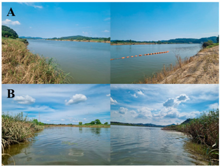 Figure 1
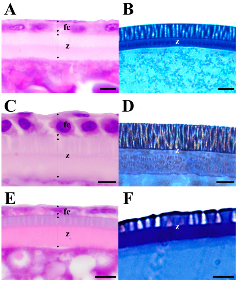 Figure 2
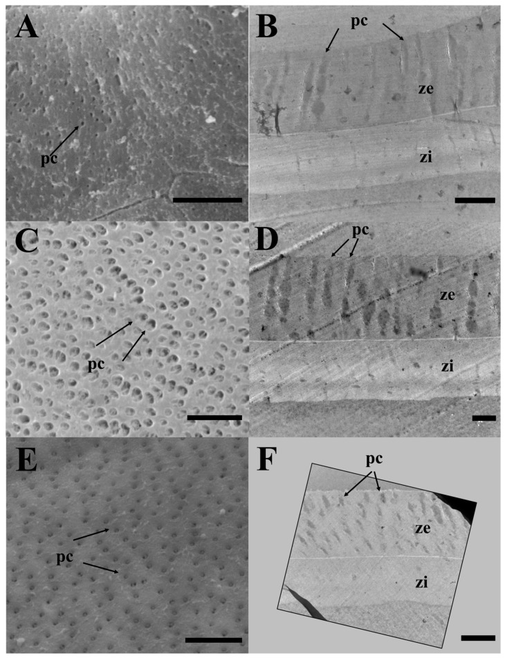 Figure 3
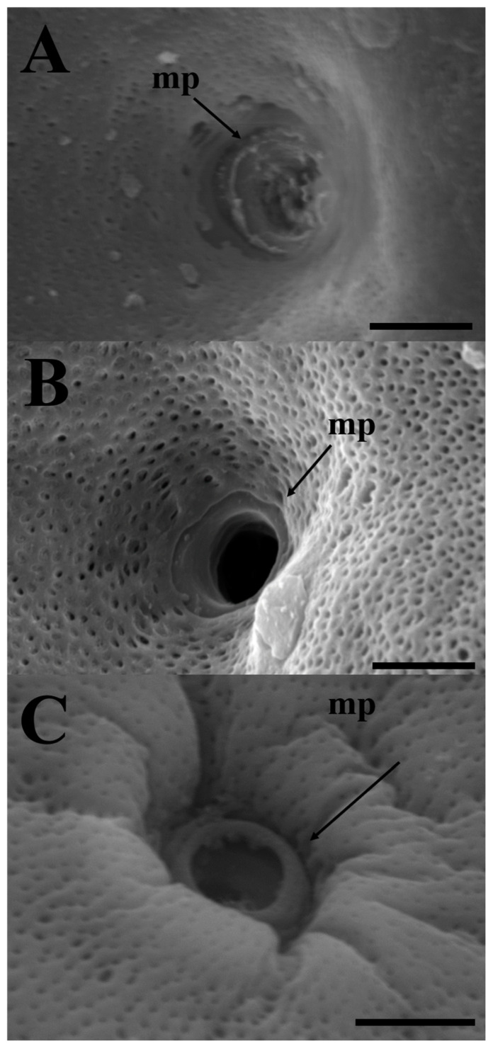 Figure 4
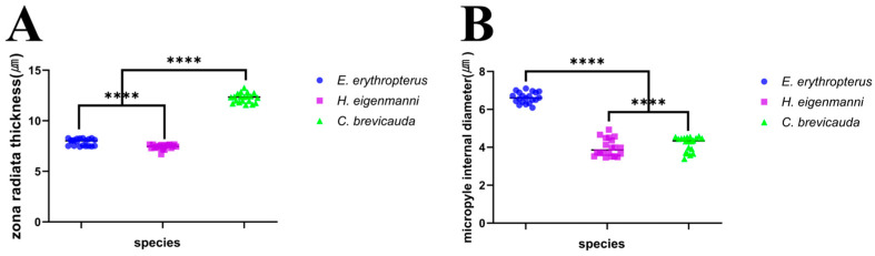 Figure 5
Peer Reviews
No public reviews on file for this paper yet. If you reviewed it on a platform where reviews are public (OpenReview, ICLR, NeurIPS, ICML), you can paste yours below so the community can read it here.
Videos
No videos yet. Explain this paper in a talk, walkthrough, or lecture? Add one.
Taxonomy
TopicsFish Ecology and Management Studies · Fish Biology and Ecology Studies · Aquaculture Nutrition and Growth
1. Introduction
The external morphology of the eggs of teleost fish varies between species, and these differences are used to create ecological and reproductive isolation. This function prevents the formation of hybrids between the different species. These differences are also important for distinguishing morphologically very similar species. In the recently developed aquaculture field, morphological studies of eggs are an important part of the development of superior species [1,2,3]. The eggs of teleosts are surrounded by a non-cellular egg envelope, the zona radiata (ZR), which is usually divided into two or three layers [4,5,6,7,8]. The inner layer is composed of a thick fibrous material, and the outer layer of a thin, highly electron-dense material [3,9]. These ZR serve to protect against physical impacts and chemical penetration from the external environment, and their structure and thickness are species-specific and influenced by habitat and spawning grounds [10,11,12].
The subfamily Cultrinae of the family Cyprinidae has approximately 18 genera and 80 species known worldwide, mostly inhabiting water systems in East Asia, including China, Mongolia, Russia, Taiwan, and Korea [13,14,15]. Four species, namely Erythroculter erythropterus, Culter brevicauda, Hemiculter eigenmanni, and Hemiculter leucisculus, from three genera, Erythroculter, Culter, and Hemiculter, have been reported to inhabit Korea [16,17]. E. erythropterus has recently been introduced to the Nakdonggang River and other rivers, showing a nationwide distribution, while C. brevicauda is only found in the Yeongsangang and Nakdonggang Rivers, but its population is declining as a result of the distribution of E. erythropterus and H. eigenmanni throughout Korea, and a very narrow range of populations living only in the estuaries of the Hangang River system is currently H. leucisculus. However, they are carnivorous in their feeding preferences, which can cause ecological disturbances when they leave their original habitats [17,18]. Since the recent introduction of H. leucisculus for aquaculture purposes in Central Asia along the Caspian Sea coast, its population has rapidly increased, causing significant effects on freshwater ecosystems [19,20,21]. In Korea, it has been reported that E. erythropterus and Opsariichthys uncirostris amurensis have invaded the Nakdonggang River from their original habitat, adversely affecting freshwater ecosystems, such as the largemouth bass Micropterus salmoides and bluegill Lepomis macrochirus, which are alien species [22]. Furthermore, in Korea, they are divided into two species (E. erythropterus and C. brevicauda) and are distinguished by morphological differences, such as carination on the abdomen and scale size. Internationally, they are not widely recognized. E. erythropterus is recognized in China and Taiwan under four different genera and scientific names, namely Chanodichthys erythropterus, C. erythropterus, E. erythropterus, and Culterichthys erythropterus, and C. brevicauda is recognized as C. alburnus, C. brevicauda, and E. adokii [13,14,23,24,25]. Moreover, C. brevicauda has been protected in Korea because its population and habitat are declining. Therefore, although they are morphologically distinct, they are accepted as synonyms, and extensive taxonomic research is required. From these points of view, Cultrinae species are causing economic and ecological damage in Korea. Firstly, the anthropogenic introduction of E. erythropterus into the habitat of endangered C. brevicauda has reduced the population of C. brevicauda and established it as the top predator in each stream, much like largemouth bass. Secondly, there is still global confusion regarding the scientific names. This study aimed to investigate the preservation of C. brevicauda populations in the face of the spread of E. erythropterus. While H. eigenmanni is clearly distinguished by morphological differences, E. erythropterus and C. brevicauda are not yet taxonomically recognized (=synonym). Although the differences in morphology are minimal, they do exist and can be used as a basis to support this argument. Therefore, we compared their oocyte structures to identify differences between species.
2. Materials and Methods
Gravid females of three species were collected using a triangle net from the Geumgang and Yeongsangang Rivers from May to July 2022 (spawning season)—E. erythropterus (36°18′14″ N, 126°55′18″ E, Hoam-ri, Gyuam-myeon, Buyeo-gun, Chungcheongnam-do, Republic of Korea) in Geumgang River, and Culter brevicauda and Hemiculter eigenmanni (35°0″2′ N, 126°41″6′ E, Godong-ri, Geumcheon-myeon, Naju-si, Jeollanam-do, Republic of Korea) in Yeongsangang River (Figure 1). The bottom structures were modified according to the Cummins [26] method using mud (M: <0.1 mm), sand (S: 0.1–2 mm), pebble (P: 2–64 mm), cobble (C: 64–256 mm), boulder (B: >256 mm), and rock (R: large stone and bedrock), and the required ratio was obtained by visual examination. The permission to catch C. brevicauda, an endangered species, was granted by the Ministry of Environment, Republic of Korea (April 2022, license number: No. 2022-22). The study followed the Guide for the Care and Use of Laboratory Animals (2011), provided by the National Institutes of Health, USA. All experimental procedures were performed under the supervision of the Institutional Animal Care and Use Committee of the Chonbuk National University, Republic of Korea. All the experiments were performed under MS-222 anesthesia, and all efforts were made to minimize pain in the animals.
After anesthetization with MS-222, samples for light microscopy analysis were obtained by removing the ovaries and analyzing the most mature eggs among the cells in the ovarian cavity. Samples for electron microscopy and toluidine staining were obtained by artificially inseminating the most mature eggs released by pressing the abdomen of the fish with male sperm (n = 20). The extracted eggs were fixed in 10% neutral buffered formalin, dehydrated using a graded ethanol series (60–100%), and cleared in xylene. The samples were embedded in wax (Paraplast, Leica, Nußloch, Germany), and 5 μm sections were deparaffinized and stained with Harris’s hematoxylin and eosin [27]. For photographs and evaluation of the ZR, a light microscope (AX 10, Carl Zeiss, Jena, Germany) was used with AxioVision (LE REL 4.5, Carl Zeiss, Jena, Germany). The oocyte size utilized in each study method was measured by selecting a total of 20 mature eggs from all 3 species and measuring their diameter. For toluidine staining, fixed tissues were dehydrated, and blocks were prepared using an Epon mixture (Epon 812, EMS, Hatfield, PA, USA). Epon blocks were sectioned at 0.8 μm using an ultramicrotome (Reichert Ultracut S, Leica, Nußloch, Germany) and stained with 1% toluidine blue. The samples were examined using a light microscope AX10 and analyzed with AxioVision 4.5.
For scanning electron microscopy (SEM), the fragments were fixed with 2.5% glutaraldehyde in 0.1 M phosphate buffer at pH 7.2. A post-fixation procedure was performed using 1.0% osmium tetroxide in the same buffer. After dehydration in a graded ethanol series (60–100%) and drying to a critical point using tert-butyl alcohol, the dried samples were coated with osmium tetroxide using a plasma coater (HPC-1SW, Vacuum Device Inc., Tokyo, Japan) and then filmed with an SEM instrument (S-300N, Hitachi, Tokyo, Japan) operating at 15 kV. For transmission electron microscopy (TEM), tissues that were fixed and dehydrated as described for the SEM were embedded in an Epon 812 mixture. The fragments were observed using a TEM device (H-7650, Hitachi, Tokyo, Japan) operating at 100 kV. The experimental values are expressed as the mean ± standard error of the mean. GraphPad Prism (version 9.0; GraphPad Inc., San Diego, CA, USA) and one-way analysis of variance (ANOVA) were used for multiple comparisons, followed by Dunnett’s test. Statistical significance was set at p < 0.001.
3. Results
Our analysis using the Cummins [26] method revealed that the substrate of the Geumgang River inhabited by E. erythropterus was composed of 80% sand, 10% mud, and 10% boulders, while the substrate of the Yeongsangang River inhabited by C. brevicauda and H. eigenmanni was more homogenous with 60% sand and 40% mud, and the habitats of the two rivers were relatively similar. As is typical of large rivers, the flow velocity is low (Figure 1). The oocyte sizes utilized in the analysis were 1.17 ± 0.09 mm for E. erythropterus, 1.08 ± 0.06 mm for C. brevicauda, and 0.79 ± 0.04 mm for H. eigenmanni. The egg size was similar between E. erythropterus and C. brevicauda, with H. eigenmanni being the smallest.
All three species (E. erythropterus, C. brevicauda, and H. eigenmanni) had non-structural forms of egg envelopes on the outer surface; no special structure other than the pore canal was identified on the outer side, and these eggs were non-adherent. The light microscopic analysis of cross-sections of mature eggs revealed striped ZR stained with eosin and 1% toluidine blue. Furthermore, no structure was identified between the outer surface of egg envelopes and the follicular cell layer. In this process, the toluidine-stained oocytes were fertilized eggs; therefore, the follicular cell layers were not identified (Figure 2).
The SEM analysis of the surface of mature eggs revealed a large number of pore canals in the egg envelopes—83 for E. erythropterus, 75 for C. brevicauda, and 58 for H. eigenmanni per 10 μm^2^ (Table 1 and Figure 3A,C,E). The TEM analysis of the ZR of the three species was similar to the results of light and SEM; multiple pore canals were identified in the ZR of E. erythropterus, C. brevicauda, and H. eigenmanni, which comprised an outer and inner layer. No other structures were identified on the outer surface of the egg envelopes (Figure 3B,D,F). The thickness of the egg envelopes was measured to be 7.89 ± 0.34 μm in E. erythropterus, 12.27 ± 0.46 μm in C. brevicauda, and 7.42 ± 0.24 μm in H. eigenmanni, and one funnel-shaped micropyle was identified at the tip of each animal pole (Figure 4). Among the three species, E. erythropterus had the largest inner diameter of the micropyle (6.62 ± 0.29 μm), followed by C. brevicauda (4.19 ± 0.39 μm), and H. eigenmanni (3.98 ± 0.46 μm) (Table 1). These results were confirmed by Dunnett’s test and one-way analysis of variance (ANOVA), which showed that the zona radiata thickness and the diameter of the micropyle for each species were significant at a confidence level of p < 0.0001. The visualization of these results using GraphPad Prism is shown in Figure 5.
4. Discussion
The outer surface of the egg envelope in fish is characterized by a variety of structures, namely non-structural, granular, villous, filamentous, saw-shaped, hillock-shaped, and fence-shaped [1,3]. These structures perform functions such as attachment, hydrostatic regulation, and embryo protection [28,29,30]. The egg envelope structures also play a role in species specificity, demonstrating a close ecological link with the habitat [31,32,33,34]. Park [1] and Choi [35] reported that the ultrastructure of an egg envelope is associated with spawning areas in fish. The non-structural form has been observed in Misgurnus anguilicaudatus, M. mizolepis, Lefua costata, Gobiobotia nakdongensis, and Microphysogobio yaluensis, which are known to have lentic habitats or spawning areas where the bottom structure comprises mud [36]. Among the Acheilognathinae that spawn in freshwater bivalves, Acheilognathus lanceolatus, A. signifer, A. koreensis, A. somjinensis, A. yamatsutae, A. majusculus and Sarcocheilichthys nigripinnis morii of the genus Sarcocheilichthys exhibit a non-structural egg envelope, which was found to be favorable for deposition in the gills of bivalves by laying fusiform, pear-shaped, and mono-oval eggs [3]. All three Cultrinae species have bottom structures composed of mud or sand and inhabit lentic waters, which is consistent with previous findings [16]. Additionally, differences in the thickness of the zona radiata of the three species (E. erythropterus, C. brevicauda, and H. eigenmanni) demonstrated species specificity.
In this study, all three species had ZR comprising two layers—an outer and inner layer. The inner layer contains several pore canals composed of microfibers, which are involved in oocyte respiration and nutrient transport [37]. In our study, we identified pore canals in all three species, and although the number varied between species, no other external structures were associated with them. A funnel-shaped micropyle with a larger outer diameter and a smaller inner diameter was observed in all three species of Cultrinae. Although the micropyle morphology of the three species was similar, there were differences observed between the species; E. erythropterus demonstrated the largest inner diameter, followed by C. brevicauda and H. eigenmanni. These micropyles are species-specific and function to prevent multi-fertilization during the spawning season and defend against invasion by sperm from other species [38,39]. Thus, C. brevicauda and H. eigenmanni, which have similar micropyle sizes, have ecological differences due to different spawning sites and times, and E. erythropterus has adapted to its environment, thus having completely different micropyle sizes. Therefore, no cases of hybridization have been reported yet between different species. Some fish have two or more micropyles, which are used as taxonomic characteristics [40,41,42]. The size of the micropyle is known to be closely related to sperm head size [43,44]. The three species in this study live in very large rivers, making it difficult to collect them with conventional nets, and the study would have been difficult to perform without equipment such as SEM and TEM instruments. If we had included the eggs of Hemiculter leucisculus in the analysis, we could have expected better results, but it is unfortunate that we could not catch them directly because they live in the Hangang River estuary. However, H. leucisculus has a similar habitat, so it is likely that it has a similar egg morphology but with different zona radiata thickness, number of pore canals, and internal size of the micropyle. Finally, we found that the egg structures of E. erythropterus and C. brevicauda, which are synonyms of scientific names because of their very similar external morphology, are different. These results can be used as a basis for species taxonomy, and we expect that they will be used as a basis for research on aquaculture and the management of invasive species using these data. This can help to manage invasive species by disturbing spawning sites, creating triploids (sterile), or interfering with natural spawning by creating artificial spawning grounds.
5. Conclusions
E. erythropterus is currently an invasive species in the Nakdonggang River, where it is a top predator that disturbs aquatic ecosystems. C. brevicauda has been ecologically declining in the Nakdonggang River and is listed as vulnerable on the IUCN Red List. H. eigenmanni is found in all water systems in Korea; therefore, the study of their egg envelope structures is expected to provide basic data for ecology, extermination, and conservation, and it is a relatively large river species that is very difficult to study. This study is, therefore, significant in that it revealed that their egg envelopes are all non-structural; the bottom structure of the stream is primarily composed of mud and sand; they live in slow-flowing water; the micropyle is funnel-shaped but differently sized in all three species; and there are differences in the thickness of the zona radiata and number of pore canals. E. erythropterus had the highest number of pore canals required for oxygenation (83), and H. eigenmanni had the lowest (58), indicating that even in areas with relatively low oxygen concentrations, E. erythropterus had a structure that favored gas exchange without the death of fertilized eggs. C. brevicauda had the thickest egg envelopes to protect the eggs from physical impact. Finally, H. eigenmanni, which lived in both E. erythropterus and C. brevicauda habitats, had the smallest micropyle, indicating that it evolved to form interspecific hybrids. These differences may be used as morphological characters to distinguish between the species of Cultrinae globally, which is currently facing problems due to several species recognized as synonyms and a lack of taxonomic research. These include E. erythropterus (named Chanodichthys erythropterus, C. erythropterus, and Culterichthys erythropterus) and C. brevicauda. This will additionally serve as a basis for aquaculture research and the management of invasive species of ecosystems in Korea.
The reference list from the paper itself. Each links out to its DOI / PubMed record.
- 1Park J.Y. A Morphological Study on the Gonad of the Species in the Family Cobitidae (Pisces: Cypriniformes) from Korea Ph.D. Dissertation Chonbuk National University Jeonju-si, Republic of Korea 1996158 p
- 2Kim S.H. Lee C.H. Ju H.S. Kim H.B. Lee Y.D. Ultrastructural variations on the micropyle of blacktip grouper, Epinephelus Fasciatus before and after artificial fertilization Korean J. Microsc.201141123128
- 3Choi S.J. Oogenesis and Ultrastructure of the Egg Envelope in 15 Korean Cyprinid Fish (Pisces, Cypriniformes) Spawning in Bivalves Ph.D. Dissertation Chonbuk National University Jeonju-si, Republic of Korea 2021122 p
- 4Anderson E. Comparative aspects of the ultrastructure of the female gamete Int. Rev. Cytol. Suppl.197441704616916 · pubmed ↗
- 5Flegler C. Electron microscopic studies on the development of the chorion of the viviparous teleost Dermogenys pusillus (Hemirhamphidae)Cell Tissue Res.197717925527010.1007/BF 00219800870211 · doi ↗ · pubmed ↗
- 6Kobayashi W. Yamamoto T.S. Fine structure of micropylar apparatus of the chum salmon egg, with a discussion of the mechanism for blocking polyspermy J. Exp. Zool.198121726527510.1002/jez.1402170213 · doi ↗
- 7Cotelli F. Andronico F. Bassi R. Brivio M. Ceccagno C. Denis-Donini S. La Rosa M.L. Lamia Donin C.L. Studies on the composition, structure and differentiation of fish egg chorion Cell Biol. Int. Rep.19861047110.1016/0309-1651(86)90046-9 · doi ↗
- 8Li Y.H. Wu C.C. Yang J.S. Comparative ultrastructural studies of the zona radiata of marine fish eggs in three genera in Perciformes J. Fish Biol.20005661562110.1111/j.1095-8649.2000.tb 00759.x · doi ↗