Concomitant Administration of Rosuvastatin and Lefleunamide in Low doses Synergize Against Complete Freunds Adjuvant (CFA)-Induced Rheumatoid Arthritis in Experimental Model
Abdel-Aziz Saeed, Mohamed El-Shafey, Gouda K. Helal, El-Sayed Akool

TL;DR
This study shows that combining low doses of rosuvastatin and lefleunamide can effectively reduce arthritis in an experimental model.
Contribution
The novel finding is that combining half-doses of rosuvastatin and lefleunamide synergistically inhibits rheumatoid arthritis.
Findings
Rosuvastatin and lefleunamide reduce inflammation and joint swelling in CFA-induced arthritis.
Combining half-doses of both drugs produces synergistic anti-arthritic effects.
Treatment improves histopathological changes and increases antioxidant activity.
Abstract
The present work was designed to examine of the potential anti-inflammatory effect of rosuvastatin (ROSV) and/or Lefleunamide (LFLU) against Complete Freunds Adjuvant (CFA)-induced arthritis in rats. The mRNA level of perxisome proliferator-activated receptor-alpha (PPAR-α) was determined using Real-time PCR. The levels of NF-κB, iNOS, IL-6, TNF-α and SOD activity were measured using ELISA. The swollen paws were measured using caliper. The GSH level was measured using colorimetric assay. The level of malondialdehyde (MDA) was determined using thiobarbituric acid reactive substances assay kit. ROSV induced the expression of PPAR-α that suppresses NF-κB as demonstrated by a strong reduction in NF-κB level in animals treated with ROSV. Also, ROSV administration reduced the levels of the inflammatory mediators IL-6 and TNF-α. In addition, iNOS and MDA content as well as expression of…
Genes, proteins, chemicals, diseases, species, mutations and cell lines named across the full text — each resolved to its canonical identifier and authoritative record.
Click any figure to enlarge with its caption.
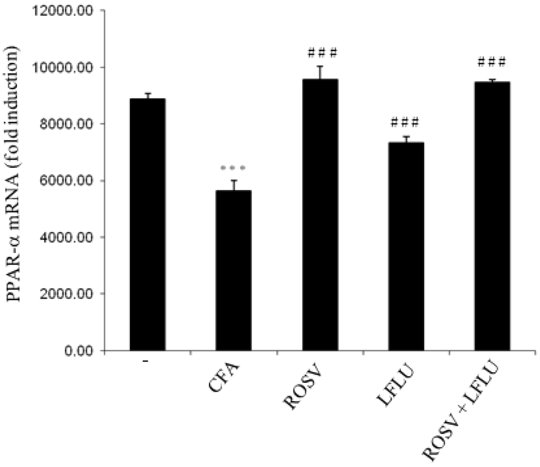 Figure 1
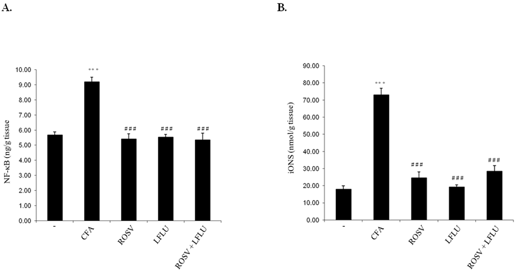 Figure 2
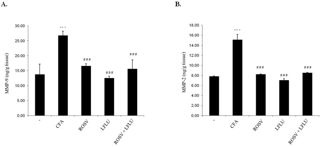 Figure 3
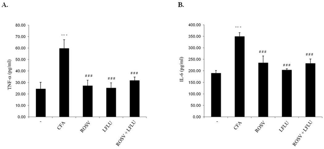 Figure 4
 Figure 5
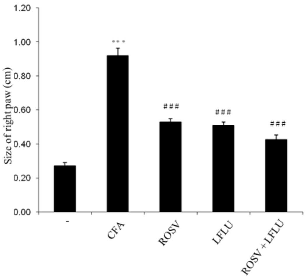 Figure 6
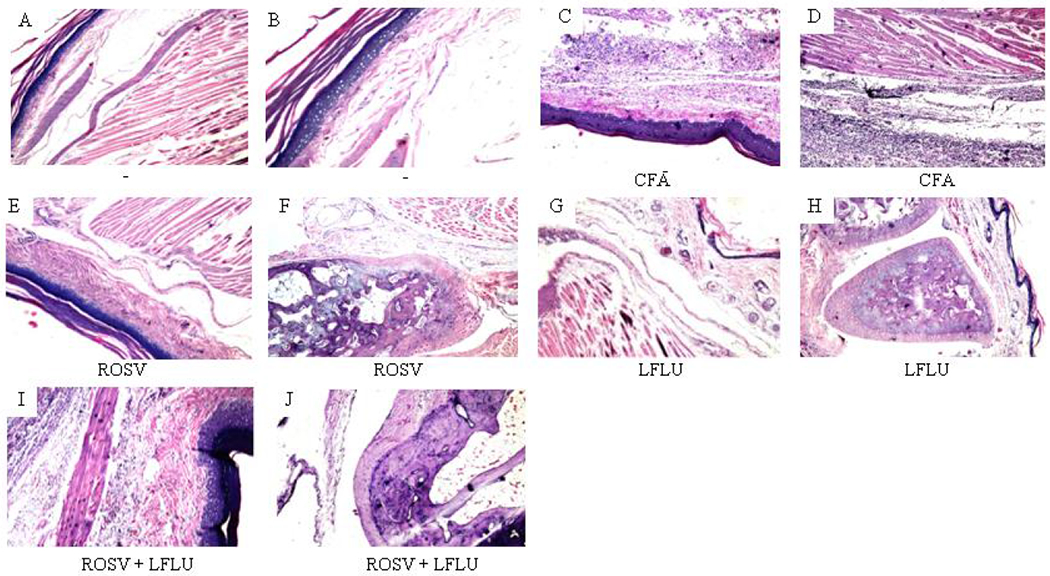 Figure 7
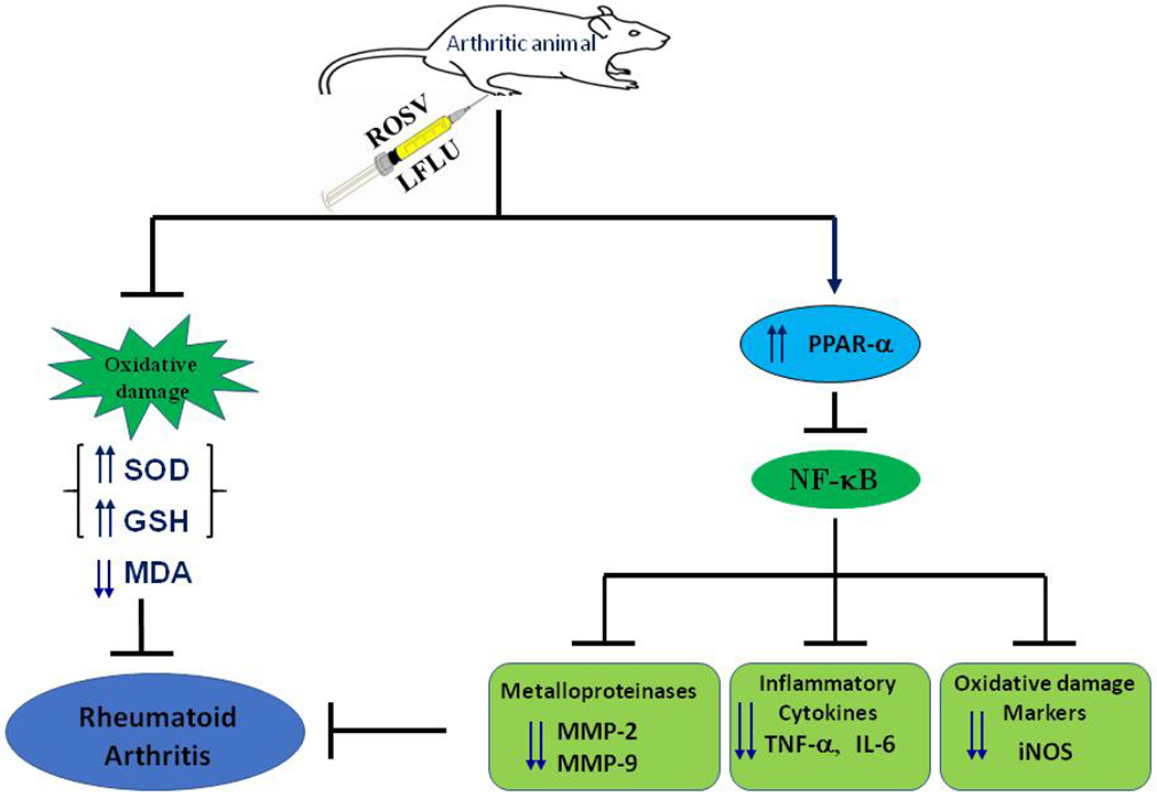 Figure 8
Peer Reviews
No public reviews on file for this paper yet. If you reviewed it on a platform where reviews are public (OpenReview, ICLR, NeurIPS, ICML), you can paste yours below so the community can read it here.
Videos
No videos yet. Explain this paper in a talk, walkthrough, or lecture? Add one.
Taxonomy
TopicsCytokine Signaling Pathways and Interactions · Synthesis and biological activity · Peroxisome Proliferator-Activated Receptors
INTRODUCTION
Rheumatoid arthritis (RA) is a chronic disease manifested by joint pain, tenderness and swelling [1]. Many studies reported the role of autoimmune phenomenon in the development of RA [2,3]. Activation of B-cells has been shown to release IgM antibody against IgG; this molecule is called rheumatoid factor (RF). The immune complexes (IgG and IgM) trigger inflammatory destruction to the synovium and collagen [2,3]. It has been shown that macrophages activation releases several cytokines which play an important role in joint tissues damage [2,3]. The destruction of the cartilage has been shown to be due to matrix metalloproteinases (MMPs) activity, produced by activated macrophages and fibroblasts in response to inflammatory cytokines such as interleukin-1 (IL-1) and tumor necrosis factor-α (TNF-α) [4]. Nuclear Factor Kappa B (NF-κB) is highly activated at sites of inflammation in variety of diseases and can control the production of proinflammatory cytokines, MMPs, adhesion molecules, inducible nitric oxide synthase (iNOS) and cyclooxygenase-2 (Cox-2) [5]. The peroxisome proliferator-activated receptors (PPARs) form a subfamily of the nuclear receptor superfamily. There are three isoforms: PPAR γ, PPAR α, and PPAR δ. The PPARs are ligand dependent transcription factors that control the transcription of different target genes through the binding to specific peroxisome proliferator response elements (PPREs) in enhancer sites of regulated genes. PPARs are expressed in immunological cell types such as monocyte/macrophages, lymphocyte, and dendritic cells. The three isoforms have been shown to inhibit the production of many inflammatory mediators and cytokines [6]. PPAR-α agonists have been shown to modulate inflammation by inhibiting cytokine (TNF-α, interleukins) production in a PPAR-α dependent manner [7–10]. Statins have been shown to induce PPAR-α expression in stimulated endothelial cells, macrophages, and hepatocytes [11]. Several drugs are usually used to manage RA like analgesics, disease-modifying anti-rheumatic drugs (DMARDs), non-steroidal anti-inflammatory drugs (NSAIDs), corticosteroids and immunosuppressive agents. Also, TNF-α and IL-1β antagonists have been used in management of RA patients [12,13]. However, the uses of these agents are usually associated with several adverse effects. Recently, researchers are directed towards the discovery of safe and effective drugs for the long-term use. Kleemann and his colleagues [14] reported that, the anti-inflammatory effect of rosuvastatin (ROSV) is mediated via peroxisome proliferator-activated receptors (PPARs) signaling-pathway through suppression of NF-κB mediated-target gene activation especially TNF-α, IL-6, adhesion molecules and iNOS. Furthermore, it has been reported that combination of atrovastatin with prednisolone produced a better results than in either remedy alone against Freunds adjuvant induced-arthritis in rats [15]. Interestingly, it has been demonstrated that inhibitors of HMG-CoA reductase may protect joints and peri-articular bones of experimental animals against experimental arthritis progression [16]. Leflunomide (LFLU) which belongs to DMARD was licensed for use in rheumatoid arthritis in 1998. It interferes with the production of inflammatory cytokines by T-cell via inhibition of NF-κB activity required for inflammatory cytokines expression [17]. It inhibits also the production of proinflammatory TNF-α and interleukin 1β [18]. In addition, LFLU has the ability to inhibit COX-2 enzyme at the site of inflammation [19]. Therefore, the present work was designed to examine first, the potential antiinflammatory effect of either ROSV or LFLU [standard DMARD] against Complete Freunds Adjuvant (CFA)-induced arthritis in rats. Second the potential anti-inflammatory effect of both ROSV and LFLU when given together in half doses against CFA-induced arthritis in rats.
MATERIALS AND METHODS
Animals
2.1
Female Wistar albino rats weighing 150-200 g were provided from Nile Co., Cairo, Egypt. The rats were housed in a 12h dark/light cycle animal facility with controlled humidity and constant temperature. A standard diet and water were supplied ad libitum. For adaptation, the rats were kept for one week under observation before the experimental study.
Materials
2.2
Complete Freunds Adjuvant (CFA) was purchased from Sigma-Aldrich Co. (St. Louis, MO, USA). Rosuvastatin was obtained from AstraZeneca Pharmaceuticals, Egypt. Lefleunamide was obtained from Multi Apex Pharmaceuticals, Egypt. Reduced glutathione (GSH), superoxide dismutase (SOD), and thiobarbituric acid reactive substances (TBARS) assay kits were purchased from Bio-diagnostic Co. (Giza, Egypt). Tumor necrosis factor-α (TNF-α) and interleukin-6 (IL-6) ELISA kits were purchased from Abcam Inc., (Cambridge, MA, USA). Inducible NO-synthase (iNOS) was purchased from EIAab Science Co., Ltd., China. Nuclear factor kappa-B (NF-κB), matrix-metalloproteinase-2 (MMP-2) and matrix-metalloproteinase-9 (MMP-9) were purchased from Cloud-Clone Corp., Houston, TX, USA.
Experimental Design
2.3
The rats were randomly divided into five groups, 8 rats in each. The control group (first group) was given the vehicle (0.5% Sodium carboxymethyl cellulose). The second group received 0.4ml CFA (SC) in right hind paw for 12 days divided in three doses to induce rheumatoid arthritis [20]. After induction of rheumatoid arthritis for 12 days, the third group were administered ROSV on day 13 in a dose of 10 mg/kg/day [21] for 28 days after the last injection of CFA. Also, after induction of rheumatoid arthritis for 12 days, the fourth group received LFLU on day 13 in a dose of 10 mg/kg/day [22] for 28 days after the last injection of CFA. On day 13, after induction of rheumatoid arthritis for 12 days, the fifth group was treated with ROSV in a dose of 5 mg/kg/day [23] plus LFLU in a dose of 5 mg/kg/day [24,25] for 28 days after the last injection of CFA. At the end, blood was collected for measurement of IL-6 and TNF-α levels. Afterwards, animals were sacrificed by cervical dislocation. Ankle, paw and knee joints were dissected immediately after death, washed with ice-cold phosphate buffered saline (PBS), and kept at −20°C for biochemical analysis. Paws specimens were kept in 10% neutral-buffered formal saline for histopathological analysis.
Assessment of NF-κB Level
2.4
The NF-κB level in joint tissue was assessed by enzyme-linked immunosorbent assay (ELISA) as previously described [26].
Assessment of iNOS Level
2.5
The iNOS level in joint tissue was detected by enzyme-linked immunosorbent assay (ELISA) according to the manufacturers instructions (EIAab Science Co.,Ltd., China).
Determination of Serum IL-6 Level
2.6
The serum level of IL-6 in was measured by enzyme-linked immunosorbent assay (ELISA) according to the manufacturers instructions (Abcam Inc., Cambridge, MA, USA).
Determination of Serum TNF-α Level
2.7
The serum level of TNF-α was detected by enzyme-linked immunosorbent assay (ELISA) according to the manufacturers instructions (Abcam Inc., Cambridge, MA, USA).
Measurement of Lipid Peroxides
2.9
The level of malondialdehyde (MDA) content in joint tissue was assessed according to the manufacturers instructions (Bio-diagnostic Co., Giza, Egypt) as previously described [26]. Briefly, the determination of TBARS calculated as MDA is based on the reaction of MDA with TBARS. The absorbance of the resultant pink color was determined at 534 nm spectrophotometrically. The MDA content in joint tissue samples was determined by comparison with the predetermined MDA standard curve.
Measurement of SOD Activity
2.10
The SOD activity in joint tissue was determined as previously described [27].
Assessment of GSH Content
2.11
The GSH content in joint tissues was assessed as previously described [26]. Briefly, GSH determination depends on the reduction of Ellmans reagent by the SH group in GSH to produce yellow product which can be measured spectrophotometrically at 405 nm.
Real-time PCR
2.12
The level of PPAR-α mRNA in joint tissue was detected using Real-time PCR system as previously described [28]. Briefly, total RNA was extracted from joint tissue according to the manufacturers instructions of the total RNA extraction kit and the RNA was subjected to reverse transcription. The following primers were used:
PPAR-α: 5′-ACGATGCTGTCCTCCTTGATG-3′ (forward),
5′- GCGTCTGACTCGGTCTTCTTG-3′ (reverse)
GAPDH: 5′-CCATTCTTCCACCTTTGATGCT-3′ (forward),
5′-TGTTGCTGTAGCCATATTCATTGT-3′ (reverse)
Real time PCR was done as follows: one initial step at 95°C for 10 min followed by 45 cycles at 95°C for 10 seconds, 55°C for 1 min and 72°C extension step for 5 seconds. The mRNA level was determined as fold change from the GAPDH level.
Measurement of Paw Size
2.13
The swollen paws were periodically examined (up to 28 days) in each paw from the ankle using caliper as previously described [29].
Histopathological Investigation
2.14
Histopathological examination was done as previously described [27].
Statistical Analysis
2.15
Results are expressed as means ± SD. One way ANOVA followed by Tukey-Kramer as a post-hoc test was used to analyze statistical significance among groups. P-values below 0.05 were considered as indication for statistically significant differences between groups compared.
RESULTS
ROSV and/or LFLU Upregulate PPAR-α mRNA Transcription in Joint Tissue of Arthritic Animals
3.1
As demonstrated in Fig. 1, induction of rheumatoid arthritis with CFA significantly reduced PPAR-α expression on mRNA level as compared to control. However, treatment of rats with either ROSV or LFLU (standard DMARD) significantly induced PPAR-α expression in joint tissue of arthritic animals. Concomitant administration of ROSV and LFLU in half doses significantly induced PPAR-α expression.
ROSV and/or LFLU Attenuate NF-κB and iNOS Expression in Experimental Model of RA Induced by CFA
3.2
Treatment of rats with either ROSV or LFLU significantly attenuated NF-κB expression induced by CFA in arthritic group (Fig. 2A). In addition, treatment of animals with ROSV in combination with LFLU in half doses significantly reduced NF-κB expression in arthritic animals (Fig. 2A). In addition, iNOS expression was highly reduced in arthritic animals treated with either ROSV or LFLU (Fig. 2B). Also, concomitant use of ROSV and LFLU in half doses significantly inhibited iNOS expression in joint tissue of arthritic rats (Fig. 2B).
ROSV and/or LFLU Downregulate the Expression of MMP-9 and MMP-2 in Joint Tissue of Arthritic Animals
3.3
As demonstrated in Fig. 3, the expression of MMP-9 and MMP-2 were highly induced in joint tissue of arthritic animals (Fig. 3A, 3B). On the other hand, treatment of arthritic rats with either ROSV or LFLU significantly inhibited the expression of MMP-9 and MMP-2 in joint tissue of arthritic animals (Fig. 3A, 3B). Concomitant use of ROSV and LFLU in half doses significantly reduced the expression of MMP-9 and MMP-2 in arthritic animals (Fig. 3A, 3B).
ROSV and/or LFLU Attenuate TNF-α and IL-6 Expression in Joint Tissue of Arthritic Animals
3.4
The level of TNF–α and IL-6 were highly increased in serum of arthritic animals (Fig. 4A, 4B). On the other hand, treatment of arthritic rats with either ROSV or LFLU significantly reduced the level of TNF-α and IL-6 (Fig. 4A, 4B). Concomitant use of ROSV and LFLU in half doses significantly reduced the level of TNF-α and IL-6 (Fig 4A, 4B).
ROSV and/or LFLU Inhibit Lipid Peroxidation in Joint Tissue of Arthritic Animals
3.5
The by-product of lipid peroxidation MDA was highly increased in joint tissue of arthritic animals (Fig. 5A). On the other hand, administration of either ROSV or LFLU significantly reduced MDA in joint tissue of arthritic animals (Fig. 5A). Concomitant use of ROSV and LFLU in half doses significantly reduced the MDA level in joint tissue of arthritic rats (Fig. 5A).
ROSV and/or LFLU Increase GSH Content and SOD Activity in Joint Tissue of Arthritic Animals
3.6
As shown in Fig. 5B, SOD activity in arthritic animals was significantly reduced. However, administration of either ROSV or LFLU significantly increased SOD activity in arthritic rats. Concomitant use of ROSV and LFLU in half doses significantly increased SOD activity in arthritic animals. Also, GSH content in joint tissue of arthritic animals was highly reduced (Fig. 5C). On the other hand, treatment of arthritic rats with either ROSV or LFLU significantly increased the GSH content in joint tissue of arthritic animals (Fig. 5C). Concomitant use of ROSV and LFLU in half doses significantly increased GSH content in arthritic animals (Fig. 5C).
ROSV and/or LFLU Decrease Paw Size in Arthritic Animals
3.7
Administration of CFA significantly induced paw in joint tissue as compared to control (Fig. 6). However, paw size was highly reduced in arthritic animals treated with either ROSV or LFLU (Fig. 6). Concomitant use of ROSV and LFLU in half doses significantly reduced paw size in arthritic animals (Fig. 6).
ROSV and/or LFLU Improved the Histopathological Alterations Induced by CFA in Joint Tissue
3.8
In contrast to control (Fig. 7A, 7B), treatment of rats with CFA significantly induced acanthosis in the epidermal layer associated with massive infiltration of inflammatory cells and aggregation in the subcutaneous tissue as well as the musculature (Fig. 7C and 7D). Interestingly, no histopathological alteration in the skin layers, subcutaneous tissue and musculature (Fig. 7E) was detected in arthritic rats treated with ROSV. Only mild degeneration was detected in the articular cartilaginous surface (Fig. 7F). Also, no histopathological alteration was detected in the skin layers, subcutaneous tissue, musculature (Fig. 7G), articular cartilaginous surface and synovial membrane (Fig. 7H) in arthritic animals treated with LFLU. Most importantly, only little hperkeratosis and mild acanthosis in the epidermis associated with few inflammatory cells infiltration in the deep dermis, subcutaneous tissue and musculature (Fig. 7I) as well as few degeneration in the cartilaginous articular surface (Fig. 7J) were detected in arthritic animals treated with both ROSV and LFLU in half doses.
DISCUSSION
Management of RA is a major health problem till now. The use of classical drugs in treatment of RA is limited due to their low safety. Therefore, it was interesting to find a new drug with high ability against inflammation and more safe. In the present study, CFA-induced rheumatoid arthritis model was used as it shares the human disease in various signs and symptoms [30]. In this study, induction of rheumatoid arthritis with CFA was found to be associated with a clear reduction in PPAR-α expression. Interestingly, treatment of rats with either ROSV or LFLU (standard DMARD) significantly induced PPAR-α expression in joint tissue of arthritic animals. Most importantly, concomitant administration of ROSV and LFLU in half doses significantly induced PPAR-α expression. This agree with previous study reported that ROSV has anti-inflammatory activity in PPARs-dependent manner [14]. Also, it has been shown that ROSV upregulates the expression of PPAR-α in vitro [31]. Interestingly, this increase in PPAR-α expression was associated with a significant reduction in NF-κB expression in arthritic animals treated with either ROSV or LFLU. These data are in line with previous study demonstrated that the antiinflammatory characters of ROSV may be attributed to its ability to inhibit NF-κB activity [4]. Most importantly, concomitant use of ROSV and LFLU in half doses significantly reduced NF-κB expression which plays an important role in the transcription of proinflammatory cytokines, MMPs, and iNOS [5]. Previously, it has been reported that the anti-inflammatory activity of statin may be attributed to its ability to inhibit iNOS expression [32,33]. In line with this study, iNOS expression was significantly reduced in arthritic animals treated with either ROSV or LFLU. Interestingly, concomitant use of ROSV and LFLU in half doses significantly inhibited iNOS expression in joint tissue of arthritic animals indicating that ROSV as well as LFLU has the ability to exert cells protection against CFA-induced nitrosative stress via inhibition of iNOS expression and subsequent reduction in NO level. Furthermore, the expression of MMP-9 and MMP-2 were highly induced in joint tissue of arthritic animals. In harmony with previous findings [34,35], treatment of arthritic rats with ROSV significantly reduced the expression of MMP-9 and MMP-2 in joint tissue. Also, LFLU administration significantly reduced the expression of MMP-9 and MMP-2 in arthritic animals. Importantly, administration of ROSV in combination with LFLU in half doses significantly reduced the expression of MMP-9 and MMP-2 in arthritic rats. Previously, it has been shown that TNF-α plays an important role in rheumatoid synovitis [36]. In the present findings, the serum level of TNF-α was highly increased in arthritic animals. Interestingly, TNF-α level was highly decreased in arthritic animals treated with either ROSV or LFLU. Similar findings were obtained in other studies [37,38]. Most importantly, concomitant use of ROSV and LFLU in half doses significantly decreased the TNF-α level. Also, IL-6 has been shown to display proinflammatory characters that are thought to be involved in the pathogenesis of RA [39]. The present work demonstrates that IL-6 level in serum of arthritic animals was highly increased. In agreement with other findings [35,40,41], administration of ROSV significantly decreased the serum IL-6 level in arthritic animals. The serum level of IL-6 was also decreased in arthritic rats treated with LFLU. Interestingly, concomitant use of ROSV and LFLU in half doses significantly decreased the serum IL-6 level in arthritic animals. The connection between oxidative stress and chronic inflammation is well known [42]. Oxidative stress is a condition in which the level of reactive oxygen species (ROS) increases overtime either by an increase in their production, decrease in the endogenous antioxidants and/or the combination of both [43]. Lipid peroxidation usually affects cell integrity [44]. The increase in the by-product of lipid peroxidation MDA in joint tissue of arthritic rats may reflect the oxidative stress. The present work shows that the by-product of lipid peroxidation MDA was highly increased in joint tissue of arthritic animals. Interestingly, administration of either ROSV or LFLU significantly reduced lipid peroxidation in joint tissue of arthritic animals. Most importantly, concomitant use of ROSV and LFLU in half doses significantly reduced lipid peroxidation in joint tissue of arthritic animals. These data are in line with previous finding demonstrated that ROSV has the ability to inhibit lipid peroxidation induced by piroxicam in gastric, liver, and kidney tissues [45]. Furthermore, SOD activity in arthritic animals was significantly reduced. In line with previous findings [46,47], administration of either ROSV or LFLU significantly increased SOD activity in arthritic group. Most interestingly, concomitant use of ROSV and LFLU in half doses significantly increased SOD activity in arthritic rats. Also, GSH content in joint tissue of arthritic animals was highly reduced. Interestingly, treatment of arthritic rats with either ROSV or LFLU significantly increased the GSH content in joint tissue of arthritic rats. These data are in agreement with previous findings [35,48]. Most importantly, concomitant use of ROSV and LFLU in half doses significantly increased GSH content in arthritic rats indicating that ROSV as well as LFLU has the ability to restore the antioxidant capacity in joint tissue of arthritic animals via enhancement of GSH content and SOD activity resulting in a great protection against oxidative stress induced by CFA. Furthermore, the present study shows that treatment of animals with CFA significantly induced paw in joint tissue compared with control. Importantly, paw size was significantly reduced in arthritic animals treated with either ROSV or LFLU. Most interestingly, concomitant use of ROSV and LFLU in half doses significantly reduced paw size in arthritic rats. Moreover, administration of either ROSV or LFLU as well as concomitant use of ROSV and LFLU in half doses significantly improved histopathological alteration in joint tissue of arthritic animals.
CONCLUSION
Our findings demonstrate that administration of either ROSV or LFLU or ROSV+LFLU (in half doses) inhibits RA in experimental model induced by CFA via induction of PPAR-α and subsequent inhibition of NF-κB resulting in a clear reduction in the inflammatory mediators (IL-6, TNF-α) and the matrix metalloproteinases (MMP-9, MMP-2) as well as iNOS expression (Fig. 8). Furthermore, oxidative stress was highly reduced in arthritic animals treated with either ROSV or LFLU or ROSV+LFLU (in half doses) as indicated by an increase in the endogenous antioxidants (SOD, GSH) and a clear reduction in the byproduct of lipid peroxidation MDA (Fig. 8). Taken together, this reduction in the inflammatory mediators and the balance between ROS production and the endogenous antioxidant defense system that was restored in joint tissues by either ROSV or LFLU or ROSV+LFLU (in half doses) are translated into a clear reduction in the size of right paw and improvement in the histopathological changes in arthritic animals. Finally, these data may support the concept of using the PPAR-α agonist ROSV as a valuable adjuvant in RA therapy. Furthermore, the use of both drugs (ROSV+LFLU) in half doses to manage RA may give similar effects that are usually obtained with the full doses and reduces the side effects that are usually caused by the full doses of these drugs. Further clinical studies are warranted to examine such an effect in human subjects.
The reference list from the paper itself. Each links out to its DOI / PubMed record.
- 1Smolen JS, Aletaha D, Koeller M, Weisman MH, Emery P. New therapies for treatment of rheumatoid arthritis. Lancet. 2007;370:1861–1874.17570481 10.1016/S 0140-6736(07)60784-3 · doi ↗ · pubmed ↗
- 2Snaith ML. ABC of rheumatology: Gout, hyperuricaemia, and crystal arthritis. BMJ. 1995;310:521–524.7888900 10.1136/bmj.310.6978.521PMC 2548885 · doi ↗ · pubmed ↗
- 3Guo Q, Wang Y, Xu D, Nossent J, Pavlos NJ, Xu J. Rheumatoid arthritis: Pathological mechanisms and modern pharmacologic therapies. Bone Res. 2018;6:15.29736302 10.1038/s 41413-018-0016-9PMC 5920070 · doi ↗ · pubmed ↗
- 4Okamoto H, Iwamoto T, Kotake S, Momohara S, Yamanaka H, Kamatani N. Inhibition of NF-kappa B signaling by fenofibrate, a peroxisome proliferator-activated receptor-alpha ligand, presents a therapeutic strategy for rheumatoid arthritis. Clin Exp Rheumatol. 2005;23:323–330.15971419 · pubmed ↗
- 5Tak PP, Firestein GS. NF-κB: A key role in inflammatory diseases. J Clin Invest. 2001;107:7–11.11134171 10.1172/JCI 11830 PMC 198552 · doi ↗ · pubmed ↗
- 6Ricote M, Glass CK. PPA Rs and molecular mechanisms of transrepression. Biochim Biophys Acta. 2007;1771:926–935.17433773 10.1016/j.bbalip.2007.02.013PMC 1986735 · doi ↗ · pubmed ↗
- 7Majdalawieh A, Ro HS. PPAR gamma 1 and LXR alpha face a new regulator of macrophage cholesterol homeostasis and inflammatory responsiveness, AEBP 1. Nucl recept signal. 2010;8:e 004.20419060 10.1621/nrs.08004 PMC 2858268 · doi ↗ · pubmed ↗
- 8Chinetti G, Fruchart JC, Staels B. Peroxisome proliferator-activated receptors (PPA Rs): nuclear receptors at the crossroads between lipid metabolism and inflammation. Inflamm Res. 2000;49:497–505.11089900 10.1007/s 000110050622 · doi ↗ · pubmed ↗