Author Correction: Cerebral vascular amyloid seeds drive amyloid β-protein fibril assembly with a distinct anti-parallel structure
Feng Xu, Ziao Fu, Sharmila Dass, AnnMarie E. Kotarba, Judianne Davis, Steven O. Smith, William E. Van Nostrand

Abstract
Genes, proteins, chemicals, diseases, species, mutations and cell lines named across the full text — each resolved to its canonical identifier and authoritative record.
Click any figure to enlarge with its caption.
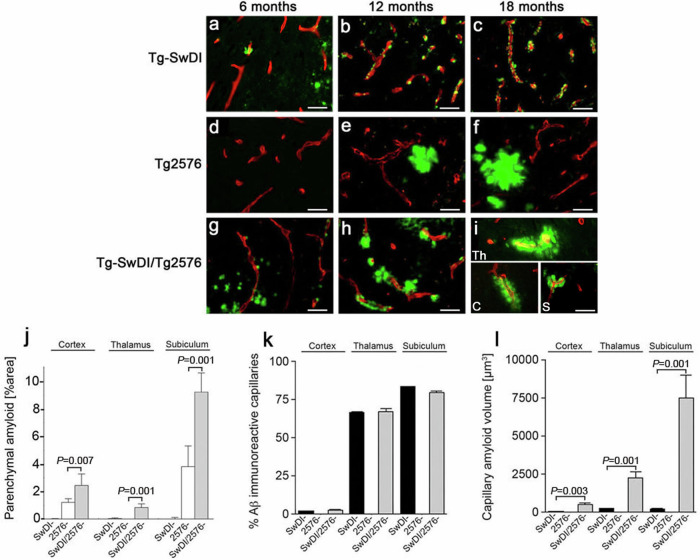 Figure 1
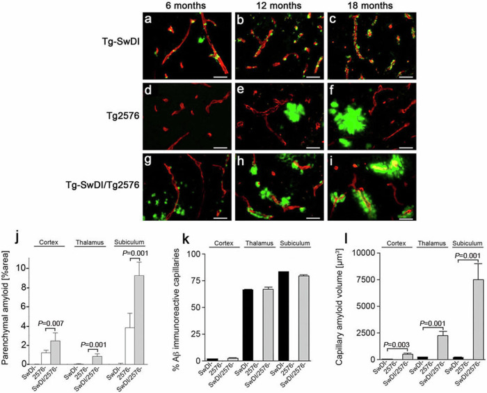 Figure 2
Peer Reviews
No public reviews on file for this paper yet. If you reviewed it on a platform where reviews are public (OpenReview, ICLR, NeurIPS, ICML), you can paste yours below so the community can read it here.
Videos
No videos yet. Explain this paper in a talk, walkthrough, or lecture? Add one.
Taxonomy
TopicsAlzheimer's disease research and treatments · Prion Diseases and Protein Misfolding · Advanced Glycation End Products research
Correction to: Nature Communications 10.1038/ncomms13527, published online 21 November 2016
The original version of this Article contained an error in Fig. 4a and Fig. 4i, and in the figure legend for Fig. 4.
In Fig. 4a, the representative image in this panel showing amyloid pathology in Tg-SwDI mice at 6 months of age was inadvertently duplicated from Fig. 4 of a previous publication by the same group^1^.
In Fig. 4i, the representative image of capillaries with enlarged amyloid deposits that were observed in 18 months old bigenic Tg-SwDI/Tg2576 mice was an aggregate of representative amyloid laden vessels from three different brain regions used for the quantitative measures presented in panels (j–l) of Figure 4, but this was not demarcated on the figure panel. The representative vessels presented in the original composite image in Fig. 4i were not used for the quantitative comparisons of percentage Aβ immune-positive capillaries (Fig. 4k) nor in measures of capillary amyloid volume (Fig. 4l) between the three brain regions.
The correct version of Fig. 4, with a separate representative image of amyloid pathology in Tg-SwDI mice at 6 months of age in Fig 4a, and appropriate demarcation of the three vessels and identified which brain region each vessel in Fig 4i, is shown here:
The incorrect version of Fig. 4 is here:
In addition, the correct figure legend for Figure 4 reads ‘(i) Composite image showing individual amyloid laden capillaries from the cortex (C), thalamus (Th), and subiculum (S) in the bigenic animals at 18 months.’ after ‘(g–i) bigenic Tg-SwDI/Tg2576 mice’.