A Rare Case of Single-Rooted Maxillary First Molar With Type II Canal Configuration
Hemal Bajaj, Rashmi Nair, Neetu Maurya, Aditya Patel, Saee Wazurkar

TL;DR
This paper describes a rare case of a maxillary first molar with a single root and a complex canal structure, successfully treated using advanced imaging and microscopy.
Contribution
The paper presents a rare clinical case and demonstrates the utility of CBCT and DOM in managing unusual root canal morphology.
Findings
A single-rooted maxillary first molar with Vertucci’s type II canal configuration was successfully treated.
CBCT and DOM were critical for diagnosing and managing the atypical canal morphology.
The case highlights the importance of advanced imaging in endodontic treatment planning.
Abstract
The morphological variations in roots and root canals vary greatly in multi-rooted teeth making it a challenge for accurate diagnosis and effective endodontic therapy. In addition to using technology appropriately, this article highlights how important it is to have a complete understanding of root canal morphology. With the assistance of cone-beam computed tomography (CBCT) images and a dental operating microscope (DOM), successful endodontic treatment was performed on a single-rooted maxillary first molar with Vertucci’s type II canal configuration. CBCT and DOM proved to be valuable tools for the effective diagnosis and management of this atypical morphology.
Genes, proteins, chemicals, diseases, species, mutations and cell lines named across the full text — each resolved to its canonical identifier and authoritative record.
Click any figure to enlarge with its caption.
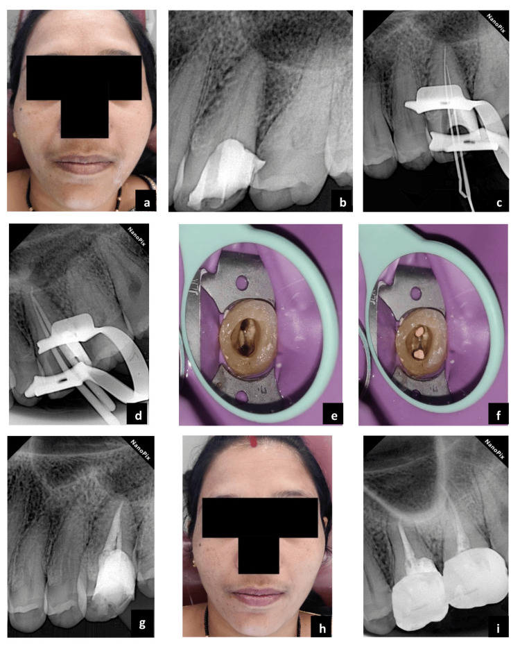 Figure 1
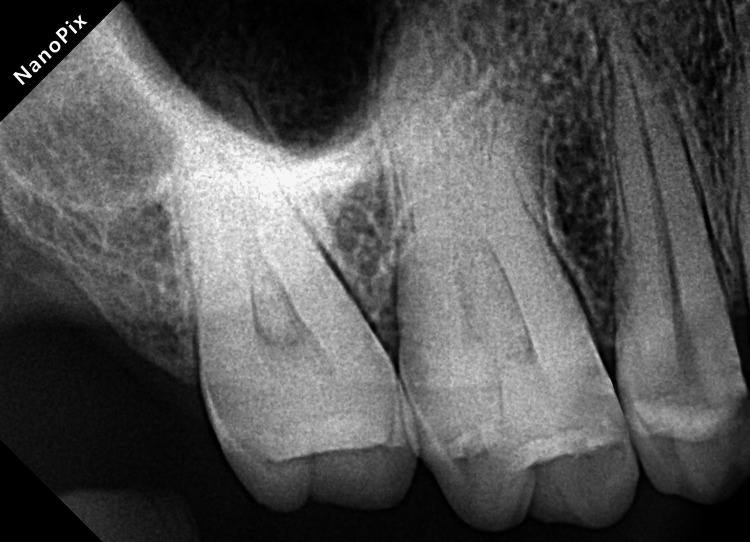 Figure 2
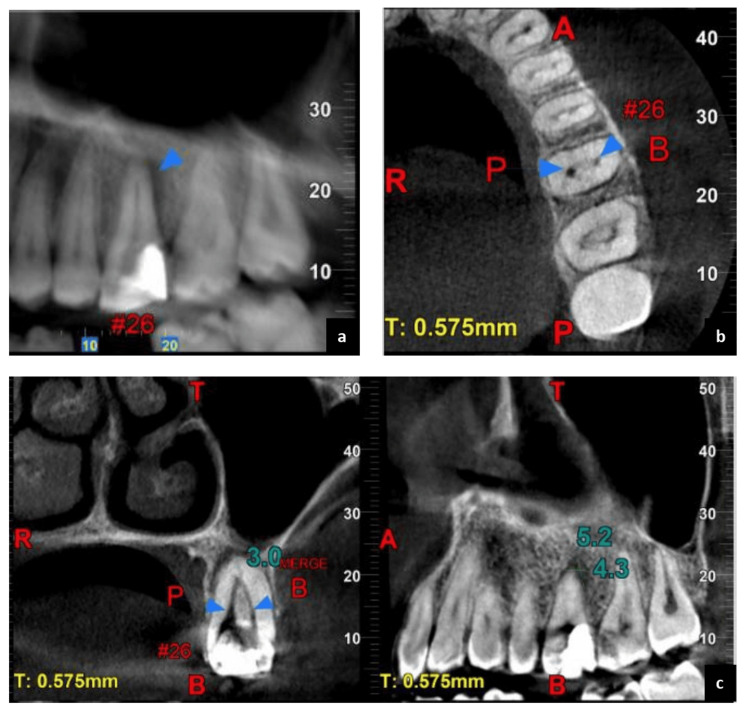 Figure 3
Peer Reviews
No public reviews on file for this paper yet. If you reviewed it on a platform where reviews are public (OpenReview, ICLR, NeurIPS, ICML), you can paste yours below so the community can read it here.
Videos
No videos yet. Explain this paper in a talk, walkthrough, or lecture? Add one.
Taxonomy
TopicsEndodontics and Root Canal Treatments · Dental Radiography and Imaging · Dental Trauma and Treatments