Evaluation of preoperative magnetic resonance imaging features and diagnostic effectiveness of grades II and III intracranial solitary fibroma
Yuncai Ran, Xiao Wang, Yong Zhang, Rui Chen, Chenchen Liu, Yunwei Ran, Weijian Wang, Xiaoyue Ma, Mengzhu Wang, Jingliang Cheng

TL;DR
This study shows that preoperative MRI features, especially ADCmin values, can help distinguish between grades II and III intracranial solitary fibrous tumors.
Contribution
The study identifies ADCmin as a significant predictor for grading ISFT using preoperative MRI, improving diagnostic accuracy.
Findings
Grade III ISFT showed more mixed T2-FLAIR and DWI signal characteristics compared to grade II.
ADCmin had an AUC of 0.805, with 74.1% sensitivity and 75.0% specificity for grading ISFT.
The model including ADCmin achieved 89.1% accuracy in differentiating grades II and III ISFT.
Abstract
To explore the value of preoperative magnetic resonance imaging (MRI) characterization of intracranial solitary fibrous tumors (ISFT) and to evaluate the effectiveness of preoperative MRI features in predicting pathological grading. This retrospective analysis comprised the clinical and preoperative MRI characterization of 55 patients with ISFT in our hospital, including 27 grade II cases and 28 grade III cases confirmed by postoperative pathology. Variables included age, sex, tumor location, cross-midline status, signal characteristics of T1-weighted imaging (T1WI), T2-weighted imaging (T2WI), T2-fluid-attenuated inversion recovery (T2-FLAIR), and diffusion‑weighted imaging (DWI), peritumoral edema, intralesional hemorrhage, focal necrosis/cystic degeneration, tumor empty vessel, maximum tumor diameter, maximum, minimum, and average values of apparent diffusion coefficient (ADCmax,…
Genes, proteins, chemicals, diseases, species, mutations and cell lines named across the full text — each resolved to its canonical identifier and authoritative record.
Click any figure to enlarge with its caption.
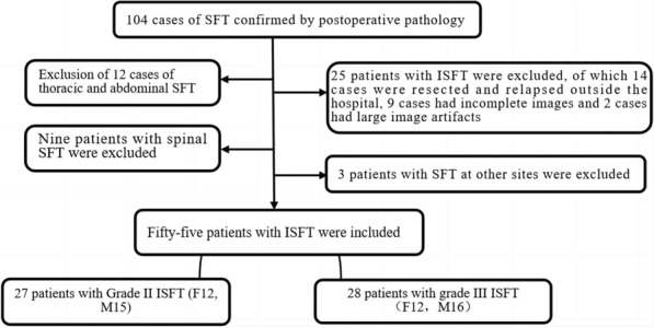 Figure 1
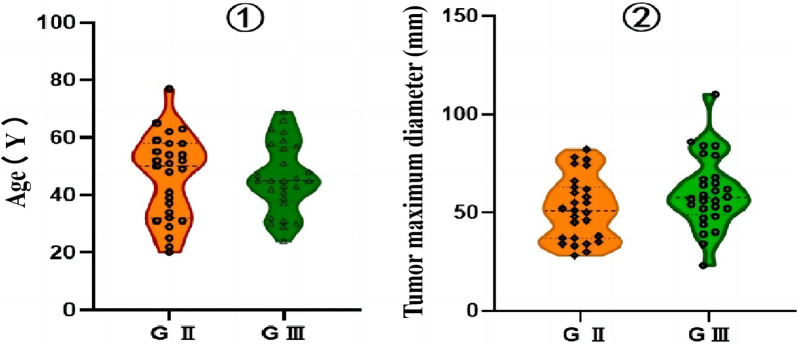 Figure 2
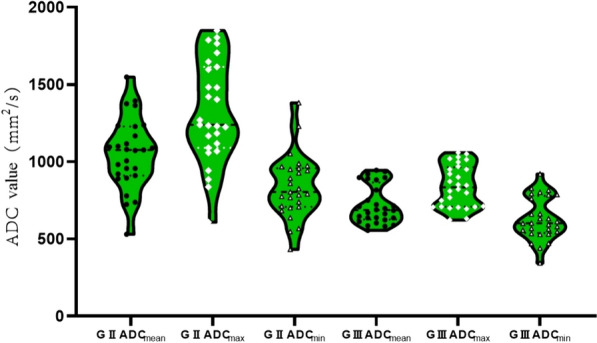 Figure 3
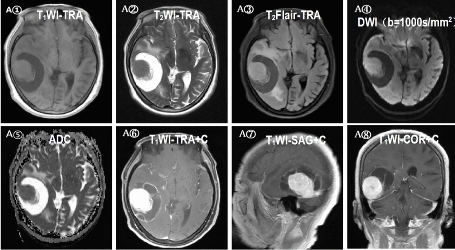 Figure 4
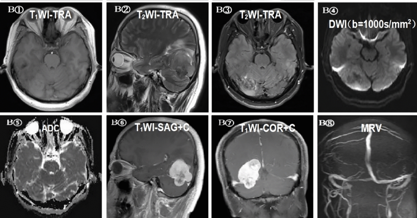 Figure 5
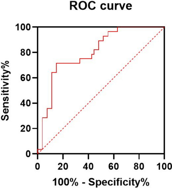 Figure 6
Peer Reviews
No public reviews on file for this paper yet. If you reviewed it on a platform where reviews are public (OpenReview, ICLR, NeurIPS, ICML), you can paste yours below so the community can read it here.
Videos
No videos yet. Explain this paper in a talk, walkthrough, or lecture? Add one.
Taxonomy
TopicsSoft tissue tumor case studies · Bone Tumor Diagnosis and Treatments · Neurofibromatosis and Schwannoma Cases