The influence of color vision deficiency on vessel visibility during colorectal endoscopic submucosal dissection and the potential advantage of red dichromatic imaging to achieve color vision barrier‐free
Akiko Ohno, Naohiko Miyamoto, Ryosuke Kaji, Takahiro Shirakawa, Moegi Watanabe, Ryutaro Sumi, Yoko Jinbo, Mitsunori Kusuhara, Jun Miyoshi, Tadakazu Hisamatsu

TL;DR
This study explores how color vision deficiency affects the ability to see blood vessels during colorectal endoscopic procedures and finds that red dichromatic imaging improves visibility for all color vision types.
Contribution
The study introduces red dichromatic imaging as a potential solution to overcome visibility challenges caused by color vision deficiency in endoscopic procedures.
Findings
Red dichromatic imaging increases color contrast between blood vessels and submucosa compared to white light imaging.
The improvement in visibility is more significant for individuals with protanopia or deuteranopia.
Red dichromatic imaging brings visibility for color-deficient individuals closer to that of people with normal color vision.
Abstract
Although color information is important in gastrointestinal endoscopy, there are limited studies on how endoscopic images are viewed by people with color vision deficiency. We aimed to investigate the differences in the visibility of blood vessels during endoscopic submucosal dissection (ESD) among people with different color vision characteristics and to examine the effect of red dichromatic imaging (RDI) on blood vessel visibility. Seventy‐seven pairs of endoscopic images of white light imaging (WLI) and RDI of the same site were obtained during colorectal ESD. The original images were set as type C (WLI‐C and RDI‐C), a common color vision. These images were computationally converted to simulate images perceived by people with color vision deficiency protanope (Type P) or deutanope (Type D) and denoted as WLI‐P and RDI‐P or WLI‐D and RDI‐D. Blood vessels and background submucosa that…
Genes, proteins, chemicals, diseases, species, mutations and cell lines named across the full text — each resolved to its canonical identifier and authoritative record.
Click any figure to enlarge with its caption.
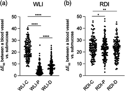 Figure 1
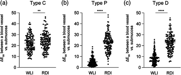 Figure 2
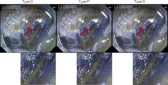 Figure 3
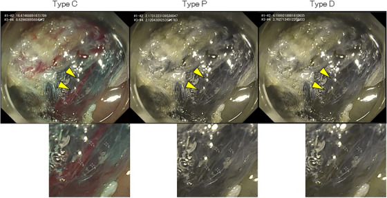 Figure 4
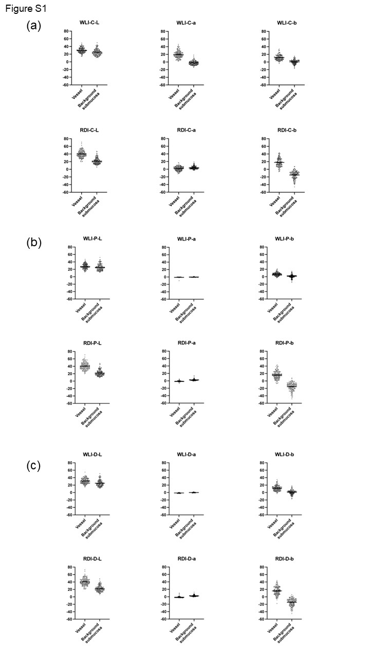 Figure 5
Peer Reviews
No public reviews on file for this paper yet. If you reviewed it on a platform where reviews are public (OpenReview, ICLR, NeurIPS, ICML), you can paste yours below so the community can read it here.
Videos
No videos yet. Explain this paper in a talk, walkthrough, or lecture? Add one.
Taxonomy
TopicsColorectal Cancer Screening and Detection · Esophageal Cancer Research and Treatment · Colorectal Cancer Surgical Treatments