Optical Coherence Tomography Characteristics for Differentiating Scars in Type 1 (Polypoidal Choroidal Vasculopathy (PCV)) and Type 2 (Classical) Macular Neovascularization (MNV) in Age-Related Macular Degeneration (AMD)
Brughanya Subramanian, Meenakshi Kumar, Parveen Sen, Rajiv Raman

TL;DR
This study uses optical coherence tomography to identify differences in scar characteristics between two types of macular neovascularization in age-related macular degeneration.
Contribution
The study identifies OCT features that can differentiate scarred stages of type 1 and type 2 macular neovascularization in AMD.
Findings
Type 2 MNV showed thinner choroid compared to type 1 MNV in most quadrants.
OCT features like outer retinal tubulations and cystoid spaces helped distinguish between the two MNV types.
Choroidal thickness differences were significant in nasal and superior quadrants within specific ranges.
Abstract
Purpose: This study aimed to assess the optical coherence tomography (OCT) characteristics for differentiating scars in the scarred stages of macular neovascularization (MNV) in age-related macular degeneration (AMD). Methods: Medical records of 20 patients, 10 in each group with type 1 and type 2 MNV, were selected for the study. Participants chosen were above 50 years of age and underwent comprehensive eye examination alongside indocyanine green angiography (ICGA), fundus fluorescence angiography (FFA), and Spectralis optical coherence tomography (SOCT) (Heidelberg Engineering, Germany), respectively. The qualitative and quantitative OCT measurements, such as the frequency of outer retinal tubulations, presence of cystoid spaces, scar area, choroid thickness, retinal thickness, presence of disorganization in retinal layers (DRIL), foveal contour, and involvement of retinal layers in…
Genes, proteins, chemicals, diseases, species, mutations and cell lines named across the full text — each resolved to its canonical identifier and authoritative record.
Click any figure to enlarge with its caption.
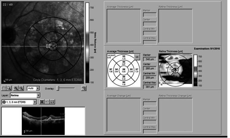 Figure 1
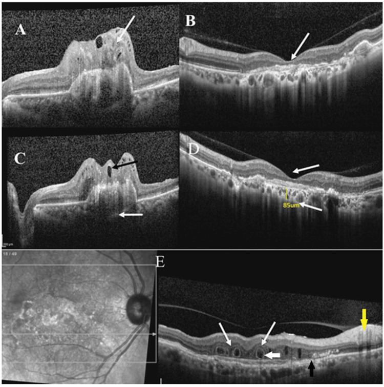 Figure 2
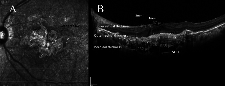 Figure 3
Peer Reviews
No public reviews on file for this paper yet. If you reviewed it on a platform where reviews are public (OpenReview, ICLR, NeurIPS, ICML), you can paste yours below so the community can read it here.
Videos
No videos yet. Explain this paper in a talk, walkthrough, or lecture? Add one.
Taxonomy
TopicsRetinal Diseases and Treatments · Retinal Imaging and Analysis · Retinal and Optic Conditions