Radiographic Features of a Strangulated Transomental Hernia
Bao H Nguyen, Aron S Mcguirt

TL;DR
An 82-year-old man with severe abdominal pain was found to have a rare strangulated transomental hernia, requiring emergency surgery to remove damaged bowel tissue.
Contribution
This case highlights the radiographic features of a rare transomental hernia causing bowel obstruction.
Findings
CT imaging showed a closed loop obstruction with anteromedial displacement of the cecum and ascending colon.
Surgery confirmed a gangrenous ileum segment strangulated by a transomental hernia.
Early recognition of radiographic signs is critical to prevent bowel ischemia.
Abstract
This is a case report of an 82-year-old male who presented with intractable and diffuse abdominal pain and had a computed tomography (CT) abdomen showing a closed loop obstruction in the right hemiabdomen with anteromedial displacement of the cecum and ascending colon. Exploratory laparotomy revealed a gangrenous segment of the ileum strangulated by a transomental hernia in the right lower quadrant. The nonviable bowel was resected, and the healthy bowel segments were anastomosed. It is important to correlate the clinical signs of bowel obstruction with radiographic findings of internal hernia to expedite surgical intervention and prevent complications of bowel ischemia.
Genes, proteins, chemicals, diseases, species, mutations and cell lines named across the full text — each resolved to its canonical identifier and authoritative record.
Click any figure to enlarge with its caption.
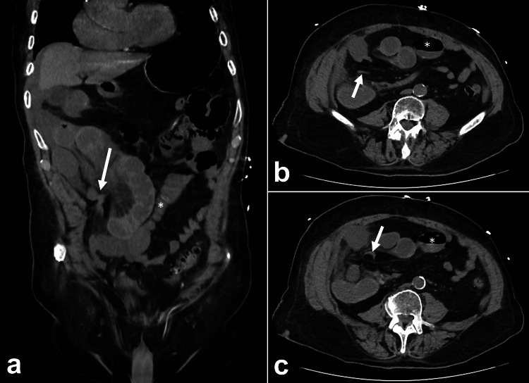 Figure 1
Peer Reviews
No public reviews on file for this paper yet. If you reviewed it on a platform where reviews are public (OpenReview, ICLR, NeurIPS, ICML), you can paste yours below so the community can read it here.
Videos
No videos yet. Explain this paper in a talk, walkthrough, or lecture? Add one.
Taxonomy
TopicsIntestinal and Peritoneal Adhesions · Hernia repair and management · Congenital Diaphragmatic Hernia Studies