Seeing is believing: a breakthrough to visualize necrosomes in the tissue
Chongbo Yang, J Magarian Blander

TL;DR
This paper introduces a new method to visualize necroptosis in tissues, helping to better understand its role in diseases like inflammatory bowel disease.
Contribution
The study presents a definitive in situ method for detecting necroptosis lesions in tissues.
Findings
The method successfully identifies necroptosis lesions in the intestinal epithelium.
The study reveals coexistence of apoptotic and necroptotic lesions in human inflammatory bowel disease.
Abstract
Detection of necroptosis in the tissue has been a long-standing roadblock in determining the disease states and pathological conditions associated with this inflammatory form of cell death. In this issue of EMBO Molecular Medicine, Chiou et al report a definitive method for necroptosis detection in situ (Chiou et al, 2024). The authors utilize this technical advance to unequivocally identify necroptosis lesions within the intestinal epithelium, and further reveal the simultaneous presence of distinct apoptotic and necroptotic lesions in human inflammatory bowel disease. JM Blander and C Yang discuss a method for necroptosis detection in situ as reported by AL Samson, JM Murphy and colleagues, in this issue of EMBO Mol Med.
Genes, proteins, chemicals, diseases, species, mutations and cell lines named across the full text — each resolved to its canonical identifier and authoritative record.
Click any figure to enlarge with its caption.
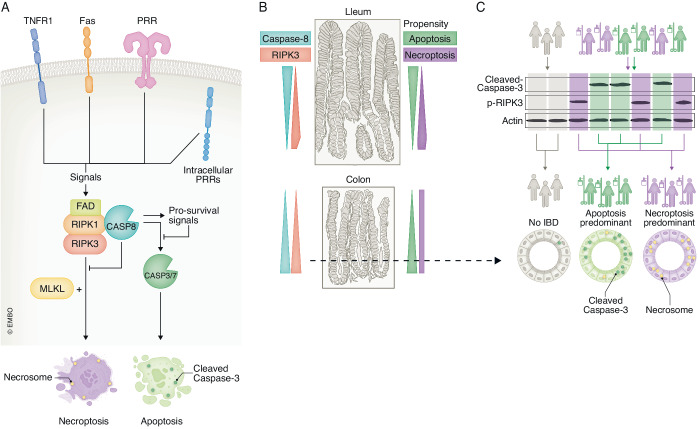 Figure 1
 Figure 2
Peer Reviews
No public reviews on file for this paper yet. If you reviewed it on a platform where reviews are public (OpenReview, ICLR, NeurIPS, ICML), you can paste yours below so the community can read it here.
Videos
No videos yet. Explain this paper in a talk, walkthrough, or lecture? Add one.
Taxonomy
TopicsCell Image Analysis Techniques · Digital Imaging for Blood Diseases