The genome sequence of a drosophilid fruit fly, Drosophila histrio (Meigen, 1830)
Darren J. Obbard, Elizabeth Everman, Anthony Bayega, Gen Morinaga

TL;DR
This paper presents the genome sequence of the drosophilid fruit fly Drosophila histrio, including a detailed assembly of its chromosomes and mitochondrial DNA.
Contribution
The paper provides the first genome assembly for Drosophila histrio, including scaffolded chromosomes and the mitochondrial genome.
Findings
The genome assembly spans 189.2 megabases and is scaffolded into 5 chromosomal pseudomolecules.
The mitochondrial genome is 16.02 kilobases in length and has been fully assembled.
Abstract
We present a genome assembly from an individual female Drosophila histrio (the drosophilid fruit fly; Arthropoda; Insecta; Diptera; Drosophilidae). The genome sequence is 189.2 megabases in span. Most of the assembly is scaffolded into 5 chromosomal pseudomolecules, including the X sex chromosome. The mitochondrial genome has also been assembled and is 16.02 kilobases in length.
Genes, proteins, chemicals, diseases, species, mutations and cell lines named across the full text — each resolved to its canonical identifier and authoritative record.
Click any figure to enlarge with its caption.
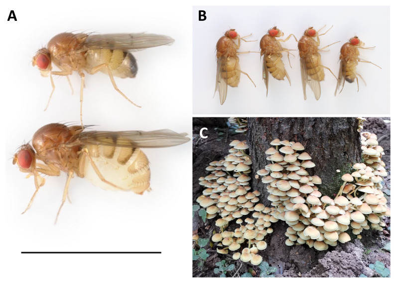 Figure 1
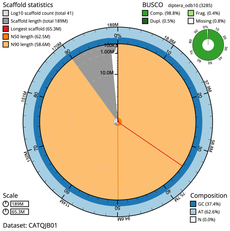 Figure 2
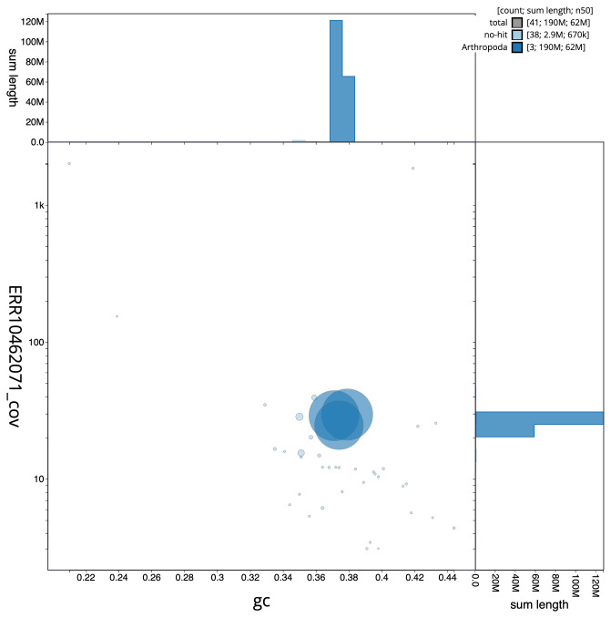 Figure 3
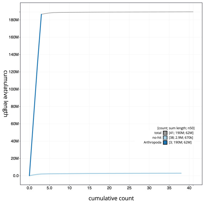 Figure 4
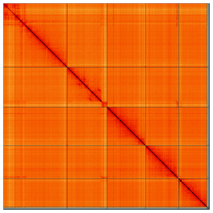 Figure 5
| Project accession data | ||
|---|---|---|
| Assembly identifier | idDroHist2.2 | |
| Species |
| |
| Specimen | idDroHist2 | |
| NCBI taxonomy ID | 198718 | |
| BioProject | PRJEB57264 | |
| BioSample ID | SAMEA12110565 | |
| Isolate information | idDroHist2, female: whole organism (DNA sequencing)
| |
| Assembly metrics
|
| |
| Consensus quality (QV) | 57.9 |
|
|
| 100.0% |
|
| BUSCO
| C:98.8%[S:98.4%,D:0.5%],F:0.4%,
|
|
| Percentage of assembly
| 99.66% |
|
| Sex chromosomes | X |
|
| Organelles | Mitochondrial genome: 16.02 kb |
|
| Raw data accessions | ||
| PacificBiosciences SEQUEL II | ERR10462071 | |
| Hi-C Illumina | ERR10466805 | |
| Genome assembly | ||
| Assembly accession | GCA_958299025.2 | |
|
| GCA_958298985.2 | |
| Span (Mb) | 189.2 | |
| Number of contigs | 440 | |
| Contig N50 length (Mb) | 1.0 | |
| Number of scaffolds | 42 | |
| Scaffold N50 length (Mb) | 36.5 | |
| Longest scaffold (Mb) | 60.84 | |
| INSDC
| Chromosome | Length (Mb) | GC% |
|---|---|---|---|
| 1 | 36.52 | 36.0 | |
| 2 | 35.08 | 38.0 | |
| 3 | 30.24 | 38.0 | |
| 4 | 25.96 | 38.5 | |
| X | 58.61 | 37.5 | |
| MT | 0.02 | 22.5 |
| Software tool | Version | Source |
|---|---|---|
| BlobToolKit | 4.1.7 |
|
| BUSCO | 5.3.2 |
|
| Hifiasm | 0.16.1-r375 |
|
| HiGlass | 1.11.6 |
|
| Merqury | MerquryFK |
|
| MitoHiFi | 2 |
|
| PretextView | 0.2 |
|
| purge_dups | 1.2.3 |
|
| sanger-tol/genomenote | v1.0 |
|
| sanger-tol/readmapping | 1.1.0 |
|
| YaHS | 1.1a.2 |
|
- —Wellcome Trust
- —UK Biotechnology and Biological Sciences Research Council
Peer Reviews
No public reviews on file for this paper yet. If you reviewed it on a platform where reviews are public (OpenReview, ICLR, NeurIPS, ICML), you can paste yours below so the community can read it here.
Videos
No videos yet. Explain this paper in a talk, walkthrough, or lecture? Add one.
Taxonomy
TopicsInsect symbiosis and bacterial influences · Genomics and Phylogenetic Studies · Insect behavior and control techniques
Species taxonomy
Eukaryota; Metazoa; Eumetazoa; Bilateria; Protostomia; Ecdysozoa; Panarthropoda; Arthropoda; Mandibulata; Pancrustacea; Hexapoda; Insecta; Dicondylia; Pterygota; Neoptera; Endopterygota; Diptera; Brachycera; Muscomorpha; Eremoneura; Cyclorrhapha; Schizophora; Acalyptratae; Ephydroidea; Drosophilidae; Drosophilinae; Drosophilini; Drosophila; Drosophila; histrio group; Drosophila histrio (Meigen, 1830) (NCBI:txid198718).
Background
Drosophila histrio Meigen,1830 is a large (4.5–6 mm) yellow-brown drosophilid ‘fruit fly’ ( Figure 1A and 1B), distantly related to the laboratory model Drosophila melanogaster. The species is broadly distributed in wooded areas across the Palaearctic, from Portugal in the west to Japan and the Kuril Islands in the far east, and from central China in the south to the north of Norway ( Bächli, 2023). It is one of around 30 British and Irish species of Drosophila ( Chandler, 2021) and, like most of its close relatives, it is a specialist fungus breeder ( Shorrocks, 1977). Adults tend to hug the forest floor and prefer decomposing and soft ephemeral fungal fruiting bodies, into which females lay a large number of small eggs ( Kimura & Toda, 1989; Toda & Kimura, 1997).
A: Male (above) and female (below) Drosophila histrio presented with a 5 mm scale bar. B: The four lab-reared siblings selected for sequencing. Sample SAMEA12110798 (centre right) was used for HiC sequencing, and sample SAMEA12110797 (centre left) was used for PacBio sequencing. C: The sulphur tuft fungus ( Hypholoma fasciculare) from which the mother of the sequenced flies was collected (Penns in the Rocks Estate, East Sussex, England; 51.093N,0.1698E).
Drosophila histrio appears notably less abundant than many other UK fungus-specialist drosophilid species ( Shorrocks, 1977), but adults have been collected from a range of fungi, such as Boletus edulis ( Morris, 2011), Polyporus squamosus ( Chandler, 2021), and Hypholoma fasciculare ( Figure 1C). Elsewhere, flies have also been reported from Pleurotus species ( Kimura & Toda, 1989), and species of Lactarius, Collybia and Russela ( Burla & Bächli, 1968). Although there are relatively few British records, D. histrio is not thought to be threatened; adults are regularly recorded in the south of the UK, with reports increasing from June to October ( GBIF, 2023).
Here we present a chromosomally complete genome sequence for Drosophila histrio, derived from the DNA of two female offspring of a wild female collected from a sulphur tuft fungus ( Hypholoma fasciculare) on the Penns in the Rocks estate, East Sussex, as part of the Darwin Tree of Life Project. This genome sequence is helping to resolve relationships among the Drosophilidae ( Kim et al., 2023), and will further build on the value of this family as a model clade for comparative genomics and molecular evolution. This project is a collaborative effort to sequence all named eukaryotic species in the Atlantic Archipelago of Britain and Ireland.
Genome sequence report
The genome was sequenced from one female Drosophila histrio ( Figure 1) reared at the Institute of Ecology and Evolution, University of Edinburgh, Scotland, UK (55.92, –3.17). A total of 45-fold coverage in Pacific Biosciences single-molecule HiFi long reads was generated. Primary assembly contigs were scaffolded with chromosome conformation Hi-C data. Manual assembly curation corrected 161 missing joins or mis-joins and removed 11 haplotypic duplications, reducing the assembly length by 0.44% and the scaffold number by 77.13%, and increasing the scaffold N50 by 15.39%.
The final assembly has a total length of 189.2 Mb in 42 sequence scaffolds with a scaffold N50 of 36.5 Mb ( Table 1). The snailplot in Figure 2 provides a summary of the assembly statistics, while the distribution of assembly scaffolds on GC proportion and coverage is shown in Figure 3. The cumulative assembly plot in Figure 4 shows curves for subsets of scaffolds assigned to different phyla. Most (99.66%) of the assembly sequence was assigned to 5 chromosomal-level scaffolds, representing 2 autosomes and the X sex chromosome. Chromosome-scale scaffolds confirmed by the Hi-C data are named in order of size ( Figure 5; Table 2). The X chromosome was identified by synteny with that of Drosophila phalerata idDroPhal2.1 (GCA_951394115.1) and has a dot chromosome fusion. The 0–15 Mb region of chromosome X is of unknown order and orientation. While not fully phased, the assembly deposited is of one haplotype. Contigs corresponding to the second haplotype have also been deposited. The mitochondrial genome was also assembled and can be found as a contig within the multifasta file of the genome submission.
Table 1.: Genome data for Drosophila histrio, idDroHist2.2.
Genome assembly of Drosophila histrio, idDroHist2.2: metrics.The BlobToolKit Snailplot shows N50 metrics and BUSCO gene completeness. The main plot is divided into 1,000 size-ordered bins around the circumference with each bin representing 0.1% of the 189,265,883 bp assembly. The distribution of scaffold lengths is shown in dark grey with the plot radius scaled to the longest scaffold present in the assembly (65,316,020 bp, shown in red). Orange and pale-orange arcs show the N50 and N90 scaffold lengths (62,479,567 and 58,608,496 bp), respectively. The pale grey spiral shows the cumulative scaffold count on a log scale with white scale lines showing successive orders of magnitude. The blue and pale-blue area around the outside of the plot shows the distribution of GC, AT and N percentages in the same bins as the inner plot. A summary of complete, fragmented, duplicated and missing BUSCO genes in the diptera_odb10 set is shown in the top right. An interactive version of this figure is available at https://blobtoolkit.genomehubs.org/view/CATQJB01/dataset/CATQJB01/snail.
Genome assembly of Drosophila histrio, idDroHist2.2: BlobToolKit GC-coverage plot.Scaffolds are coloured by phylum. Circles are sized in proportion to scaffold length. Histograms show the distribution of scaffold length sum along each axis. An interactive version of this figure is available at https://blobtoolkit.genomehubs.org/view/CATQJB01/dataset/CATQJB01/blob.
Genome assembly of Drosophila histrio, idDroHist2.2: BlobToolKit cumulative sequence plot.The grey line shows cumulative length for all scaffolds. Coloured lines show cumulative lengths of scaffolds assigned to each phylum using the buscogenes taxrule. An interactive version of this figure is available at https://blobtoolkit.genomehubs.org/view/CATQJB01/dataset/CATQJB01/cumulative.
Genome assembly of Drosophila histrio, idDroHist2.2: Hi-C contact map of the idDroHist2.2 assembly, visualised using HiGlass.Chromosomes are shown in order of size from left to right and top to bottom. An interactive version of this figure may be viewed at https://genome-note-higlass.tol.sanger.ac.uk/l/?d=J3MI3e8oQYSdTkL7Bwflrw.
Table 2.: Chromosomal pseudomolecules in the genome assembly of Drosophila histrio, idDroHist2.
The estimated Quality Value (QV) of the final assembly is 57.9 with k-mer completeness of 100.0%, and the assembly has a BUSCO v5.3.2 completeness of 98.8% (single = 98.4%, duplicated = 0.5%), using the diptera_odb10 reference set ( n = 3,285).
Metadata for specimens, barcode results, spectra estimates, sequencing runs, contaminants and pre-curation assembly statistics are given at https://links.tol.sanger.ac.uk/species/198718.
Methods
Sample acquisition and nucleic acid extraction
Drosophila histrio specimens were first-generation female progeny from a wild-collected female. The sequenced flies were reared at the University of Edinburgh (latitude 55.92, longitude –3.17), and were harvested on 2021-11-10. The mother was collected from a sulphur tuft fungus ( Hypholoma fasciculare) at Penns in the Rocks Estate, East Sussex, England (latitude 51.093, longitude 0.170) on 2021-09-07. The fly was collected and identified by Darren Obbard (University of Edinburgh), and species identification was confirmed by examination of the progeny. Flies were reared on laboratory Drosophila medium with the addition of a ~2cm ^3^ piece of commercial mushroom ( Agaricus bisporus) to encourage egg laying. Each living anaesthetised fly was placed directly into the collection tube and frozen from live at –80 °C. The sample with specimen ID SAN00002002 (ToLID idDroHist2 was used for DNA sequencing and the sample with specimen ID SAN00002003 (ToLID idDroHist3) was used for Hi-C scaffolding.
The workflow for high molecular weight (HMW) DNA extraction at the WSI includes a sequence of core procedures: sample preparation; sample homogenisation, DNA extraction, fragmentation, and clean-up. In sample preparation, the idDroHist2 sample was weighed and dissected on dry ice ( Jay et al., 2023). Tissue from the whole organism was homogenised using a PowerMasher II tissue disruptor ( Denton et al., 2023a). HMW DNA was extracted using the Manual MagAttract protocol ( Strickland et al., 2023b). HMW DNA was sheared into an average fragment size of 12–20 kb in a Megaruptor 3 system with speed setting 30 ( Todorovic et al., 2023). Sheared DNA was purified by solid-phase reversible immobilisation ( Strickland et al., 2023a): in brief, the method employs a 1.8X ratio of AMPure PB beads to sample to eliminate shorter fragments and concentrate the DNA. The concentration of the sheared and purified DNA was assessed using a Nanodrop spectrophotometer and Qubit Fluorometer and Qubit dsDNA High Sensitivity Assay kit. Fragment size distribution was evaluated by running the sample on the FemtoPulse system.
Protocols developed by the Wellcome Sanger Institute (WSI) Tree of Life core laboratory are publicly available on protocols.io ( Denton et al., 2023b).
Sequencing
Pacific Biosciences HiFi circular consensus DNA sequencing libraries were constructed according to the manufacturers’ instructions. DNA sequencing was performed by the Scientific Operations core at the WSI on a Pacific Biosciences SEQUEL II instrument. Hi-C data were also generated from whole organism tissue of idDroHist3 using the Arima2 kit and sequenced on the Illumina NovaSeq 6000 instrument.
Genome assembly, curation and evaluation
Assembly was carried out with Hifiasm ( Cheng et al., 2021) and haplotypic duplication was identified and removed with purge_dups ( Guan et al., 2020). The assembly was then scaffolded with Hi-C data ( Rao et al., 2014) using YaHS ( Zhou et al., 2023). The assembly was checked for contamination and corrected as described previously ( Howe et al., 2021). Manual curation was performed using HiGlass ( Kerpedjiev et al., 2018) and Pretext ( Harry, 2022). The mitochondrial genome was assembled using MitoHiFi ( Uliano-Silva et al., 2023), which runs MitoFinder ( Allio et al., 2020) or MITOS ( Bernt et al., 2013) and uses these annotations to select the final mitochondrial contig and to ensure the general quality of the sequence.
A Hi-C map for the final assembly was produced using bwa-mem2 ( Vasimuddin et al., 2019) in the Cooler file format ( Abdennur & Mirny, 2020). To assess the assembly metrics, the k-mer completeness and QV consensus quality values were calculated in Merqury ( Rhie et al., 2020). This work was done using Nextflow ( Di Tommaso et al., 2017) DSL2 pipelines “sanger-tol/readmapping” ( Surana et al., 2023a) and “sanger-tol/genomenote” ( Surana et al., 2023b). The genome was analysed within the BlobToolKit environment ( Challis et al., 2020) and BUSCO scores ( Manni et al., 2021; Simão et al., 2015) were calculated.
Table 3 contains a list of relevant software tool versions and sources.
Wellcome Sanger Institute – Legal and Governance
The materials that have contributed to this genome note have been supplied by a Darwin Tree of Life Partner. The submission of materials by a Darwin Tree of Life Partner is subject to the ‘Darwin Tree of Life Project Sampling Code of Practice’, which can be found in full on the Darwin Tree of Life website here. By agreeing with and signing up to the Sampling Code of Practice, the Darwin Tree of Life Partner agrees they will meet the legal and ethical requirements and standards set out within this document in respect of all samples acquired for, and supplied to, the Darwin Tree of Life Project.
Further, the Wellcome Sanger Institute employs a process whereby due diligence is carried out proportionate to the nature of the materials themselves, and the circumstances under which they have been/are to be collected and provided for use. The purpose of this is to address and mitigate any potential legal and/or ethical implications of receipt and use of the materials as part of the research project, and to ensure that in doing so we align with best practice wherever possible. The overarching areas of consideration are:
• Ethical review of provenance and sourcing of the material
• Legality of collection, transfer and use (national and international)
Each transfer of samples is further undertaken according to a Research Collaboration Agreement or Material Transfer Agreement entered into by the Darwin Tree of Life Partner, Genome Research Limited (operating as the Wellcome Sanger Institute), and in some circumstances other Darwin Tree of Life collaborators.
The reference list from the paper itself. Each links out to its DOI / PubMed record.
- 1Abdennur N Mirny LA : Cooler: Scalable storage for Hi-C data and other genomically labeled arrays. Bioinformatics. 2020;36(1):311–316. 10.1093/bioinformatics/btz 540 31290943 PMC 8205516 · doi ↗ · pubmed ↗
- 2Allio R Schomaker‐Bastos A Romiguier J : Mito Finder: Efficient automated large‐scale extraction of mitogenomic data in target enrichment phylogenomics. Mol Ecol Resour. 2020;20(4):892–905. 10.1111/1755-0998.13160 32243090 PMC 7497042 · doi ↗ · pubmed ↗
- 3Bächli G : Taxo Dros: The Database on Taxonomy of Drosophilidae. 2023; [Accessed 29 May 2023]. Reference Source
- 4Bernt M Donath A Jühling F : MITOS: Improved de novo metazoan mitochondrial genome annotation. Mol Phylogenet Evol. 2013;69(2):313–319. 10.1016/j.ympev.2012.08.023 22982435 · doi ↗ · pubmed ↗
- 5Burla H Bächli G : Beitrag zur kenntnis der schweizerischen Dipteren, inbesondere drosophila-arten. Die sich in fruchtkorpern von Hutpilzen entwickeln. Vierteljahresch Naturforschenden Gesellschaft in Zürich. 1968;113:311–336.
- 6Challis R Richards E Rajan J : Blob Tool Kit - interactive quality assessment of genome assemblies. G 3 (Bethesda). 2020;10(4):1361–1374. 10.1534/g 3.119.400908 32071071 PMC 7144090 · doi ↗ · pubmed ↗
- 7Chandler PJ : Diptera of Windsor Forest and Great Park. Dipterists Digest. 2021;28. Reference Source
- 8Cheng H Concepcion GT Feng X : Haplotype-resolved de novo assembly using phased assembly graphs with hifiasm. Nat Methods. 2021;18(2):170–175. 10.1038/s 41592-020-01056-5 33526886 PMC 7961889 · doi ↗ · pubmed ↗