Single ulcers on the tongue dorsum: differential diagnosis between paracoccidioidomycosis and squamous cell carcinoma
Claudia Rebecca Costa Cavalcante, Maria Emília Mota, José Divaldo Prado, Oslei Paes de Almeida, Christianne Soares Moreira Barbosa, Joao Adolfo Costa Hanemann, Norberto Nobuo Sugaya, Maria Stella Moreira, Fábio Abreu Alves

TL;DR
This study compares paracoccidioidomycosis and oral squamous cell carcinoma as possible causes of single ulcers on the tongue dorsum, emphasizing the need for biopsy to confirm diagnosis.
Contribution
The study highlights the differential diagnosis of rare tongue dorsum ulcers between PCM and OSCC, emphasizing the importance of biopsy.
Findings
Nine patients with single tongue dorsum ulcers were diagnosed with either PCM or OSCC.
OSCC was the main clinical hypothesis despite PCM being a significant differential diagnosis.
Biopsy is essential for accurate diagnosis and treatment planning.
Abstract
Paracoccidioidomycosis (PCM) is the leading cause of death among systemic mycoses in Brazil. On the other hand, oral squamous cell carcinoma (OSCC) is the most prevalent malignant neoplasm of the mouth. Both lesions rarely affect the tongue dorsum and may share similar clinical characteristics. This study aimed to retrieve cases of single oral ulcers diagnosed as PCM or OSCC. A cross-sectional retrospective study was conducted. All patients who had a single ulcer on dorsum of the tongue and confirmed diagnosis of PCM or OSCC were evaluated. A total of 9 patients (5 women and 4 men) were evaluated, 5 patients had OSCCs (mean age = 69,8 years old), and 4 patients PCM (mean age = 51 years old). Most of the lesions were infiltrated and indurated in the palpation exam. Duration ranged from 1 to 12 months (mean time of 5.2 months and 4.7 months for OSCC and PCM, respectively). OSCC was the…
Genes, proteins, chemicals, diseases, species, mutations and cell lines named across the full text — each resolved to its canonical identifier and authoritative record.
Click any figure to enlarge with its caption.
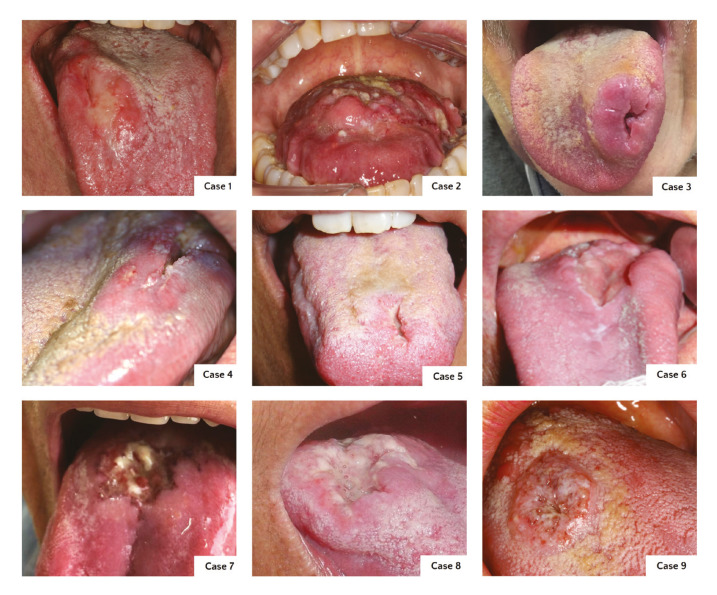 Figure 1
Peer Reviews
No public reviews on file for this paper yet. If you reviewed it on a platform where reviews are public (OpenReview, ICLR, NeurIPS, ICML), you can paste yours below so the community can read it here.
Videos
No videos yet. Explain this paper in a talk, walkthrough, or lecture? Add one.
Taxonomy
TopicsFungal Infections and Studies · Oral Health Pathology and Treatment · Viral-associated cancers and disorders
Introduction
Paracoccidioidomycosis (PCM) is an endemic and neglected disease in Brazil, caused by Paracoccidioides brasiliensis and Paracoccidioides lutzii (1,2). It is accepTable that the main if not the only form of contamination occurs through the inhalation of spores (3-5). Most patients evolve as a primary pulmonary focus, developing a chronic infection. In some cases, hematogenous and/or lymphatic propagation occurs in other locations, such as skin, adrenal glands, central nervous system, bones, and oral mucosa (6).
Oral PCM manifestations consist of multiple moriform lesions/ulcers, with the gingiva and alveolar ridge mucosa responsible for 51% of the affected sites of the mouth (7). On the other hand, Oral Squamous Cell Carcinoma (OSCC), which is responsible for 90% of oral cancer, commonly presents as a single irregular ulcer in the lateral border of the tongue. It is well known that OSCC is related to intense and prolonged exposure to tobacco and alcohol, most in men over 50 years of age. However, in the last 3 decades, the epidemiological profile has been changing, with the diagnosis of patients younger than 45 years old and lesions originating from HPV infection (8).
Although lesions affecting the dorsum of the tongue are uncommon, some studies have described cases of amyloidosis, hamartomas, leiomyomatosis, syphilis, and OSCC (9-14). An interesting case of PCM affecting the dorsum of the tongue had OSCC as the main diagnosis hypothesis (14). Thus, the present study aimed to compare cases of single oral ulcers on the tongue dorsum diagnosed as PCM or OSCC and discuss its main features.
Material and Methods
This cross-sectional retrospective study was approved by the Institutional Committee on Ethics (no 4.754.713). All cases presenting as a single ulcer on the dorsum of the tongue with a confirmed diagnosis of PCM or OSCC were evaluated. Clinical pictures (patients’ photography) were accessed to evaluate the clinical characteristics. Patients who had multiple PCM lesions or PCM/OSCC in other regions of the mouth were excluded.
Demographic data, habits, time, symptoms, diagnosis hypothesis, and stages were collected from patients’ charts.
Results
A total of 9 patients, 5 women and 4 men, were evaluated. OSCCs were diagnosed in 5 (mean age = 69,8 years old, ranging from 58-88), and PCM in 4 patients (mean age = 51 years old, ranging from 41-72. Tobacco use was reported in all cases of OSCC, and alcohol in 2. Of the PCM patients, all 4 patients were smokers, 2 were alcoholics and 1 patient denied any habits (Table 1).
Both OSCC and PCM patients presented a single ulcer on the dorsum of the tongue. Most of the lesions were infiltrated and indurated in the palpation exam, and duration ranged from 1 to 12 months (mean time of 5.2 months and 4.7 months for OSCC and PCM, respectively) (Fig. 1).
Figure 1. Oral squamous cell carcinoma (cases 1 to 5) and paracococcidioidomycosis (cases 6 to 9) on the dorsum.
Pain was reported by 3 patients with OSCC and in only one with PCM. For all cases, OSCC was the main clinical diagnosis hypothesis and syphilis and PCM were also considered in the PCM cases. Two patients with OSCC were considered with advanced stages based on TNM staging (Table 1).
Discussion
PCM is a clinical differential diagnosis of OSCC, mainly if PCM presents as a single lesion in the mouth (14,15), particularly on the dorsum of the tongue (14). Here, we are reporting additional 9 cases, 5 OSCC and 4 PCM, affecting the dorsum of the tongue. The similarity of the clinical features of both lesions is evident in the Figures here shown. All 4 cases of PCM had OSCC as the main diagnosis hypothesis, and PCM, tuberculosis, and syphilis were other options for clinical diagnosis. Moreover, some authors added histoplasmosis, leishmaniasis, syphilis, and Wegener's granulomatosis as other differential diagnoses of single oral lesions of PCM (16).
Oral mucosa is involved in 50% of the cases of PCM (2), and in general the lesions are multiple with moriform surface. Gingiva/alveolar ridge (23.2%), lips (21.7%), and buccal mucosa (15.9%) are the main locations. The tongue is involved in around 11.2% of the cases. The presence of single lesions, as in our cases, is extremely rare (7,14,16). Men are more frequently affected by PCM (19:1), between the ages of 30 and 60 years (3). Epidemiological studies indicate the rarity of PCM in women, especially during the reproductive years, which suggests a protective action of the female hormones (17). Although multiple lesions are the most common presentation of oral PCM as mentioned before, our findings shared the same features as those presented by de Oliveira Gondak, where a significant part of the patients who presented a single lesion were women (15). Therefore, further studies evaluating the association of single oral lesions with the influence of female hormones are needed.
OSCC on the dorsum tongue is extremely rare. In a Japanese study that evaluated 368 tongue SCC, only three cases affected the dorsum (0.8%) (18). In addition, such tumors had their diagnoses in advanced stage due to delay in suspecting of malignancy (18,19). Two out 5 cases of our series had advanced clinical stage. It is important to define the appropriate diagnosis of these single ulcers in the dorsum of the tongue to promptly establish adequate treatment. Both diseases, OSCC and PCM, may present similarities in the patient profile and lesion clinical features, however, the therapeutic modality is totally different. PCM is treated with prolonged antifungal medicines (1,3) and, in contrast, OSCC is treated with surgical approaches and other modalities are added in more advanced stages, such as radiotherapy and chemotherapy (20,21). Therefore, an early and correct diagnosis with histopathological analysis can prevent unnecessary invasive/radical treatment.
In summary, PCM and OSCC should be considered as diagnostic hypotheses in single and infiltrating ulcers on the dorsum of the tongue. After detailed anamnesis and clinical examination, an incisional biopsy is mandatory to define the diagnosis and appropriate treatment of single ulcers in the tongue dorsum. Finally, it is interesting to consider that our cases of PCM here described were more frequent in women, and this observation deserves to be better evaluated in future epidemiological studies.
The reference list from the paper itself. Each links out to its DOI / PubMed record.
- 1Hahn RC Rodrigues AM Terra PP Nery AF Hoffmann-Santos HDGóis HM Clinical and epidemiological features of paracoccidioidomycosis due to Paracoccidioides lutzii P Lo S Negl Trop Dis 201913 e 00074373116302810.1371/journal.pntd.0007437 PMC 6548353 · doi ↗ · pubmed ↗
- 2Singi P Rocha R Pde Carli ML Hanemann JAC Pereira AAC Coelho LFL Different DNA methylation profile is demonstrated in paracoccidioidomycosis patients without oral lesions Mycoses 201962113393150506710.1111/myc.13000 · doi ↗ · pubmed ↗
- 3Meneses-García A Mosqueda-Taylor A Morales-de la Luz R Rivera LMRG Paracoccidioidomycosis: Report of 2 cases mimicking squamous cell carcinoma Oral Surgery, Oral Medicine, Oral Pathology, Oral Radiology, and Endodontology 200294609131242445610.1067/moe.2002.129179 · doi ↗ · pubmed ↗
- 4Cordova LA Torres J Paracoccidioidomycosis. 2022 Sep 19In: Stat Pearls [Internet]Treasure Island (FL): Stat Pearls Publishing 2024 Jan.P Mid
- 5Shikanai-Yasuda MA Mendes RP Colombo AL Queiroz-Telles FQ Kono ASG Paniago AM Brazilian guidelines for the clinical management of paracoccidioidomycosis Rev Soc Bras Med Trop 201750715402874657010.1590/0037-8682-0230-2017 · doi ↗ · pubmed ↗
- 6da Silva J de Fde Oliveira HC Marcos CM Assato PA Fusco-Almeida AM Mendes-Giannini MJS Advances and challenges in paracoccidioidomycosis serology caused by Paracoccidioides species complex: an update Diagn Microbiol Infect Dis 20168487942649454110.1016/j.diagmicrobio.2015.06.004 · doi ↗ · pubmed ↗
- 7Girardi FM Scroferneker ML Gava V Pruinelli R Head and Neck Manifestations of Paracoccidioidomycosis: An Epidemiological Study of 36 Cases in Brazil Mycopathologia 2012173139442198977310.1007/s 11046-011-9488-5 · doi ↗ · pubmed ↗
- 8Hussein AA Helder M Nde Visscher JG Leemans CR Braakhuis BJ Vet HCW Global incidence of oral and oropharynx cancer in patients younger than 45 years versus older patients: A systematic review Eur J Cancer 201782115272865478510.1016/j.ejca.2017.05.026 · doi ↗ · pubmed ↗