When the Usual Becomes Unusual: A Closer Look at Mycobacterium marinum Infections
Ioannis Kyriazidis, Myrto Trakatelli, Georgia-Alexandra Spyropoulou

TL;DR
This paper discusses a case of a rare Mycobacterium marinum infection that was misdiagnosed for months and eventually treated successfully with targeted antibiotics.
Contribution
The paper highlights the diagnostic challenges and treatment approach for M. marinum infections through a detailed case study.
Findings
M. marinum infections can present as chronic, ulcerated skin lesions that resist standard treatments.
Combination therapy with minocycline and rifampicin effectively resolved the infection in this case.
Patient history and histology are crucial for diagnosing M. marinum due to its nonspecific symptoms.
Abstract
Mycobacterium marinum (M. marinum) is a slow-growing bacterium predominantly found in aquatic environments. While not highly virulent, it can cause skin and soft tissue infections, often misdiagnosed due to their indolent progression. This paper presents the case of a 42-year-old male data analyst with a chronic, ulcerated lesion on his right middle finger resulting from a minor fish tank injury. Despite multiple interventions, the lesion resisted healing for 10 months. A detailed history raised the suspicion of atypical mycobacterial infection. Despite non-diagnostic initial evaluations, combined antimicrobial therapy with minocycline and rifampicin led to complete lesion healing. Diagnosing M. marinum infection remains a challenge due to its nonspecific presentation. Key diagnostic criteria include resistance to standard antibiotics, history of exposure to aquatic environments, and…
Genes, proteins, chemicals, diseases, species, mutations and cell lines named across the full text — each resolved to its canonical identifier and authoritative record.
Click any figure to enlarge with its caption.
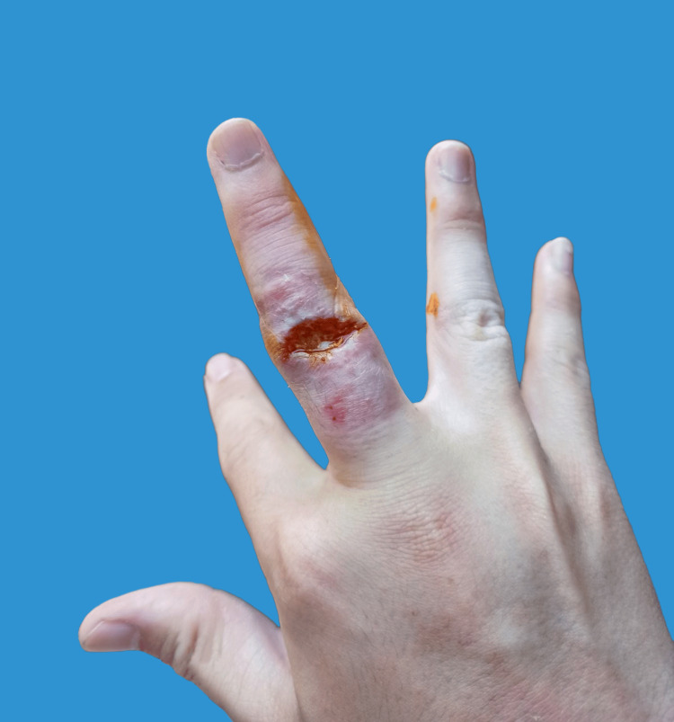 Figure 1
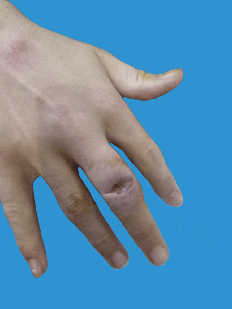 Figure 2
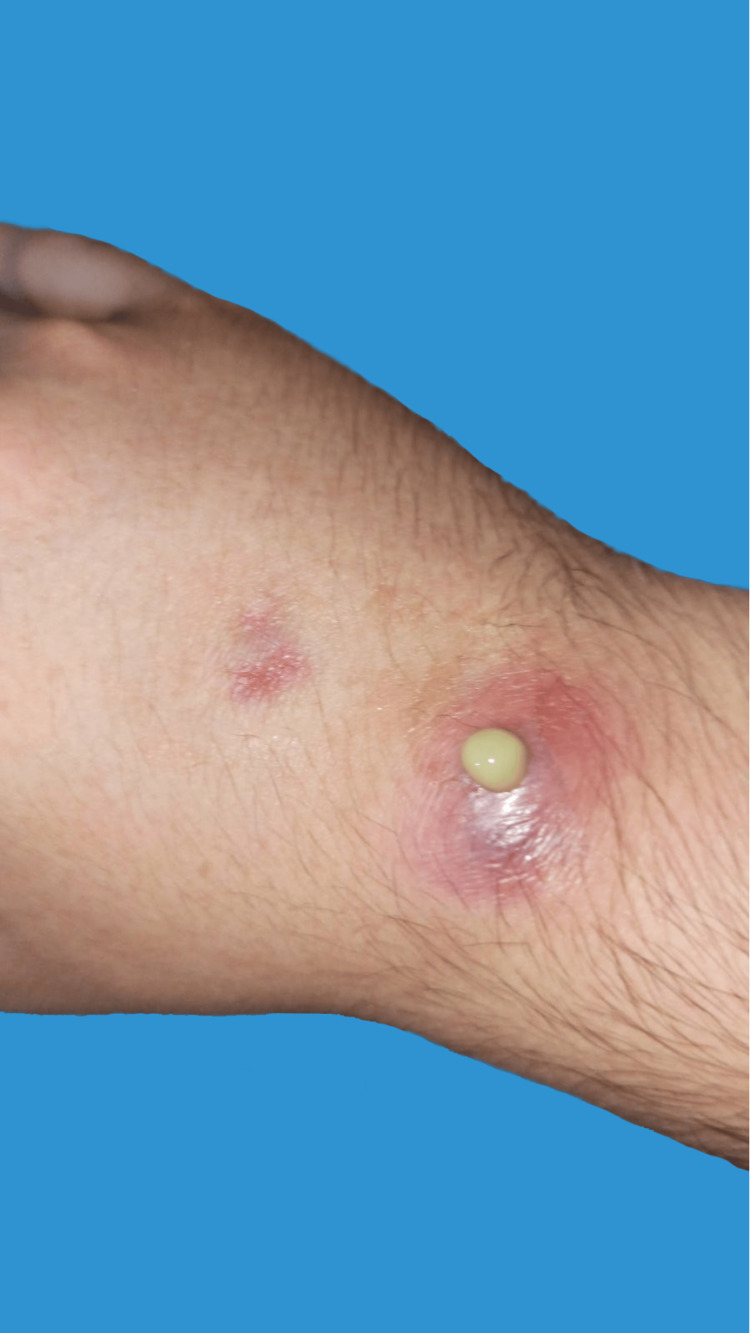 Figure 3
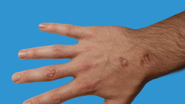 Figure 4
Peer Reviews
No public reviews on file for this paper yet. If you reviewed it on a platform where reviews are public (OpenReview, ICLR, NeurIPS, ICML), you can paste yours below so the community can read it here.
Videos
No videos yet. Explain this paper in a talk, walkthrough, or lecture? Add one.
Taxonomy
TopicsMycobacterium research and diagnosis · Tuberculosis Research and Epidemiology
Introduction
Mycobacterium marinum (hereafter M. marinum) is a slow-growing, acid-fast, environmental mycobacterium commonly found in fresh and saltwater environments. Its biology is complex and has been extensively studied. M. marinum is a facultative intracellular bacterium that can infect macrophages and other phagocytic cells, leading to a persistent infection[1].
Despite its low virulence, it can cause infections in humans, particularly in those with impaired skin barriers[2]. These typically present as skin and soft tissue infections, such as nodules, papules, and ulcers, and are often misdiagnosed as other skin conditions, such as cellulitis or pyoderma, due to their slow growth and indolent clinical course. Moreover, instances of chronic granulomatous tenosynovitis post-M. marinum infection have been documented[3]. The diagnosis is often delayed, leading to prolonged treatment and poor outcomes[1].
The primary mode of transmission is through direct skin contact with contaminated water, manifesting as cutaneous and/or subcutaneous lesions, which may be solitary or multiple, nodular, or ulcerated, predominantly observed on the extremities of the upper limbs, including fingers and hands[1]. Individuals who are at an increased risk of infection include those with a history of exposure to fresh and saltwater environments, like aquarium hobbyists and fish handlers, as well as those with compromised skin barriers, such as those with eczema or other skin conditions[4].
In addition to direct skin contact, M. marinum can also spread through lymphatic drainage, leading to the clinical appearance of linear sporotrichoid lesions[5], that can be particularly challenging to treat, as they often require prolonged courses of antimicrobial therapy and can result in significant morbidity[1].
We present a compelling case that exemplifies the challenges and considerations associated with M. marinum infections, shedding light on its diagnostic and therapeutic intricacies.
Case presentation
A 42-year-old male, right-hand-dominant data analyst, with no significant past medical history presented with a chronic, ulcerated lesion located on the dorsum of the proximal phalanx of his right middle finger. This lesion emerged around 10 months prior after the patient sustained a minor cut while cleaning a fish tank. Despite the lesion's self-dehiscence shortly after the trauma (Figure 1), it had persistently resisted healing. He had attempted multiple therapeutic interventions including topical steroid ointments, courses of oral flucloxacillin, regenerative ointments, cryotherapy, multiple wound debridements, and even a failed direct closure, all to no avail.
Ulcerative lesion over the PIPJ manifesting self-discharge of pus, captured approximately two weeks post-injury by the patient.PIPJ: proximal interphalangeal joint
Upon clinical examination, the lesion was characterized as a well-defined ulcerative defect, approximately 1 cm in diameter, surrounded by an erythematous, elevated margin (Figure 2).
Clinical presentation of the lesion during initial assessment in the outpatients' clinic. It is characterized by a well-defined ulcerative defect, approximately 1 cm in diameter. The central area displays necrotic tissue with purulent exudate, lacking surrounding erythema or warmth, indicative of the persistent nature of M. marinum infection.
The center of the lesion was necrotic with purulent exudate, but notably lacked surrounding erythema or warmth. Additionally, there were two tender erythematous nodules on the dorsum of the proximal hand and wrist that were self-discharging pus (Figure 3).
During the initial presentation, the patient was displaying two erythematous nodules expressing pus, identified as sporotrichoid lesions, on the dorsum of the proximal hand and wrist.
Motor and sensory functions of the hand and digits were intact, and no axillary lymphadenopathy was detected. Furthermore, the patient denied systemic symptoms such as fever, night sweats, or weight loss.
Radiological evaluation via MRI did not show osteomyelitis neither joint involvement but revealed soft tissue swelling. Despite the non-diagnostic outcomes from the subsequent soft tissue biopsy and wound cultures, a heightened level of suspicion for an atypical mycobacterial infection was engendered, primarily influenced by the patient's detailed hobbyist history. This led to the initiation of a three-month combined antimicrobial therapy with minocycline and rifampicin. Over this therapeutic period, the patient experienced a consistent reduction in pain and swelling. The lesion exhibited marked improvement and eventually healed completely (Figure 4) with no recorded recurrence during an eight-month follow-up period, with the patient presenting a full active range of movement to the digit and hand.
Image taken several weeks post-initiation of a prolonged course of antibiotic therapy with minocycline and rifampicin, exhibiting substantial recession of the infection and significant healing.
Discussion
Cutaneous infection due to M. marinum is relatively infrequent, and its diagnostic journey often proves elusive due to its subtle and nonspecific manifestation. Typically, there's a pronounced gap between symptom onset and etiologic agent identification[5]. The incubation period typically spans fewer than four weeks; however, it can extend significantly, with some case reports documenting periods of up to nine months[6].
A heightened diagnostic suspicion is typically anchored on a triad: a cutaneous lesion showing minimal response to standard antibiotics, a history of exposure to aquatic environments (common among aquarium enthusiasts and fish farmers)[5], and a background of potential contamination.
Previous findings indicate that cultures are positive in approximately 70-80% of cases[1,3]; thus, negative cultures in the diagnosis of M. marinum infections, though not commonplace, represent a significant challenge in clinical practice. The slow growth rate of M. marinum combined with its unique metabolic needs can contribute to suboptimal growth in standard culture media, potentially leading to false-negative results [6], as in this case. Furthermore, prior antibiotic treatments, even if ineffective for M. marinum, can further reduce the bacterial load, making culture detection even more challenging. In cases where cultures remain negative despite high clinical suspicion, next-generation sequencing (NGS) is an emerging diagnostic tool that can aid in identifying slow-growing atypical mycobacteria like M. marinum from clinical specimens[7]. NGS allows for rapid, culture-independent detection of mycobacterial DNA sequences that may be present in low abundance, providing an alternative method when routine cultures fail to yield a definitive diagnosis [1]. However, the absence of a positive culture result can create diagnostic ambiguity, which underscores the importance of maintaining a high clinical suspicion. In many scenarios, the lesions caused by M. marinum don't follow a distinct pattern.
Diagnostic clarity may be further clouded by a broad differential diagnosis that includes other non-tuberculous mycobacteria, sporotrichosis, and several non-infectious conditions[5]. Treatment modalities are influenced by guidelines and generally advocate for a combination antibiotic regimen lasting until lesion healing, followed by 1-2 additional months[1,8]. Notably, M. marinum is sensitive to a range of medications but resistant to others[8].
Thus, a synergistic approach involving histological assessment and mycobacterial studies (comprising both cultures and molecular diagnostics if available) is pivotal for its accurate identification.
Treatment regimens, like those opted for in our presented case, are typically tailored to individual patient needs, considering both lesion severity and potential underlying conditions. Surgical interventions play a pivotal role in certain cases, particularly extensive lesions[1]. The prognosis for M. marinum cutaneous infections remains optimistic, especially when treated before any deeper tissue or bone involvement [1,8]. This is clearly evidenced by the prompt treatment and favorable outcome of our reported case.
Conclusions
In light of this case and the supporting literature, it's evident that M. marinum infections, though not highly virulent, can pose significant diagnostic and therapeutic challenges. A thorough patient history, especially concerning exposure to aquatic environments, combined with a high degree of clinical suspicion, can guide clinicians towards a more accurate and timely diagnosis. This case serves as a reminder of the importance of considering atypical pathogens in the differential diagnosis of chronic skin lesions and the value of persistent and targeted antimicrobial therapy for achieving favorable outcomes.
The reference list from the paper itself. Each links out to its DOI / PubMed record.
- 1Mycobacterium marinum Microbiol Spectr Aubry A Mougari F Reibel F Cambau E 5201710.1128/microbiolspec.tnmi 7-0038-2016 PMC 1168747928387180 · doi ↗ · pubmed ↗
- 2Twenty-eight cases of Mycobacterium marinum infection: retrospective case series and literature review Infection Johnson MG Stout JE 6556624320152586982010.1007/s 15010-015-0776-8PMC 6535045 · doi ↗ · pubmed ↗
- 3Mycobacterium marinum as a cause of chronic granulomatous tenosynovitis in the hand J Infect Pang HN Lee JY Puhaindran ME Tan SH Tan AB Yong FC 5845885420071720785910.1016/j.jinf.2006.11.014 · doi ↗ · pubmed ↗
- 4Fish tank exposure and cutaneous infections due to Mycobacterium marinum: tuberculin skin testing, treatment, and prevention Clin Infect Dis Lewis FM Marsh BJ von Reyn CF 3903973720031288416410.1086/376628 · doi ↗ · pubmed ↗
- 5Mycobacterium marinum lymphocutaneous infection Dermatol Online J Bouceiro-Mendes R Ortins-Pina A Fraga A 13030252019 https://pubmed.ncbi.nlm.nih.gov/30865411/30865411 · pubmed ↗
- 6Incubation period and sources of exposure for cutaneous Mycobacterium marinum infection: case report and review of the literature Clin Infect Dis Jernigan JA Farr BM 4394433120001098770210.1086/313972 · doi ↗ · pubmed ↗
- 7Genomic characterization of nontuberculous mycobacteria Sci Rep Fedrizzi T Meehan CJ Grottola A 45258720172834563910.1038/srep 45258 PMC 5366915 · doi ↗ · pubmed ↗
- 8Treatment of Mycobacterium marinum cutaneous infections Expert Opin Pharmacother Rallis E Koumantaki-Mathioudaki E 29652978820071800125610.1517/14656566.8.17.2965 · doi ↗ · pubmed ↗