Fetal dystocia in guinea pigs: A case report
Julia Penna de Andrade, Sabrina de Morais Miranda, Camilla Faria Soares, Thaís Larissa Lourenço Castanheira, Bruno Ferrante, Marcelo Pires Nogueira de Carvalho

TL;DR
This case report details the surgical treatment of dystocia in a guinea pig, emphasizing the challenges and importance of early intervention.
Contribution
The report provides a detailed account of a rare surgical case in guinea pigs with dystocia, highlighting diagnostic and therapeutic approaches.
Findings
Dystocia in guinea pigs can be caused by large fetal size and pubic symphysis mineralization.
Surgical intervention via ovariohysterectomy successfully removed three fetuses in a dystocia case.
Postoperative care with anti-inflammatory and antibiotic treatments supported recovery, though prognosis remains guarded.
Abstract
Dystocia is a common complication in guinea pig pregnancies, presenting significant challenges in clinical management. The present case report describes the presentation, diagnosis, and surgical intervention in an 8 months old female guinea pig with dystocia. The subject is a primiparous guinea pig originating from a commercial breeder, exhibited prolonged labor with two pups, one of which was stillborn. Physical examination revealed a distended abdomen, lack of uterine contractions, signs of distress, and vulvar discharge. Radiographic and ultrasound tests confirmed obstruction due to large fetal size and mineralization of the pubic symphysis. Surgical intervention proceeded with a ventral midline approach, ovariohysterectomy and removal of three fetuses. The guinea pig recovered well from the procedure, being discharged with postoperative care, and the use of anti-inflammatory,…
Genes, proteins, chemicals, diseases, species, mutations and cell lines named across the full text — each resolved to its canonical identifier and authoritative record.
Click any figure to enlarge with its caption.
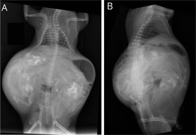 Figure 1
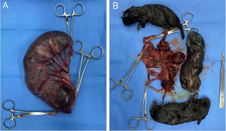 Figure 2
Peer Reviews
No public reviews on file for this paper yet. If you reviewed it on a platform where reviews are public (OpenReview, ICLR, NeurIPS, ICML), you can paste yours below so the community can read it here.
Videos
No videos yet. Explain this paper in a talk, walkthrough, or lecture? Add one.
Taxonomy
TopicsVeterinary Medicine and Surgery · Veterinary Pharmacology and Anesthesia · Veterinary Equine Medical Research
Introduction
The guinea pig (Cavia porcellus) belonging to the order Rodentia, is widely recognized as a medium-sized rodent and is popular as a non-conventional pet (Smith, 2012). Gestation in this species typically ranges between 68-72 days, with labor lasting approximately 30 minutes, interspersed with breaks of 3-7 minutes for the delivery of subsequent fetuses (Czarnecki & Adamski, 2016). Puberty is reached within 55 to 70 days, with females exhibiting their first estrous cycle by 89 days (Couto, 2006; Lapchik et al., 2017), although both male and females can reach maturity and fertility before 2 months of age (Genzer et al., 2023). Some authors advocate for the initiation of breeding before 6-7 months of age, primarily to mitigate the risk of dystocia, attributed to the stiffening of the pubic symphysis, which interferes with relaxation during birth. Studies have reported varying outcomes based on parental age, with adults showing higher fetal delivery rates, while juveniles present an elevated survival rate (Genzer et al., 2023). Litter size ranges from one to eight fetuses, with an average of 2.75 offsprings (Couto, 2006), each pup weighing between 60-100g (Kaiser et al., 2010). The primary causes of dystocia include pelvic symphysis fusion in unbred females and large fetal size, leading to a poor prognosis due to late detection, often requiring caesarean section. The objective of the case report herein is to review the caesarean section and ovariohysterectomy performed on an 8 –months old guinea pig pregnant with five fetuses, along with the subsequent follow-up treatment.
Case description
An 8 –months old female guinea pig weighing 880g was scheduled for an appointment due to dystocia. The subject came from a commercial breeder and this was her first litter. The tutor reported a labor duration of 12 hours, during which the guinea pig delivered two pups, one live and one stillborn. The following vital parameters were assessed: pulse rate at 150 beats per minute, respiratory rate at 88 breaths per minute, 39°C temperature, and 10% dehydration, with a glucose level of 108mg/dL. Physical examination revealed a significantly distended abdomen, firm on palpation, with no evident uterine contractions. The tutor had administered calcium and oxytocin, obtained without veterinary prescription and in unknown dosages, prompted by advice from social media. Upon failure of this unguided treatment, the guinea pig was brought in for a veterinary appointment, exhibiting signs of distress, prostration and pain, with a bloody and mucoid vulvar discharge. Given the severity of the situation, surgical intervention was deemed necessary, despite specific rates of surgical necessity in guinea pig dystocia cases not being well-documented when compared to dogs and cats (Gilson, 2003). Imaging tests were requested to assess fetal distress and determine the surgical intervention demand. Blood samples could not be collected prior to surgery because the in-hospital laboratory was already closed.
Radiographic examination revealed the presence of three fetuses with mineralized skeletons (Figure 1), indicating a physical obstruction likely attributable to fetus size and pubic symphysis mineralization. Ultrasonography further confirmed the lack of fetal viability, evidenced by the absence of heart rate and uterine contractions, consistent with prolonged labor and fetus distress. Given the ineffectiveness of prior clinical treatments and the impracticability of pelvic delivery due to fetal size and labor duration, surgical intervention was deemed necessary. Prior to surgery, the guinea pig was premedicated with enrofloxacin 5% (10mg/kg IM), meloxicam 0.2% (0.5mg/kg IM), dipyrone 500mg/mL (25mg/kg PO), and fluid therapy with ringer (20ml/kg SQ). Anesthesia was induced with diazepam (0.5mg/kg IM), ketamine (5mg/kg IM), morphine (2mg/kg IM), and maintained trans-surgically with inhaled sevoflurane (2%).
Radiography of Cavia porcellus (female, 8 months and 880g) presenting dystocia. (A) Ventro-dorsal projection showing three large, mineralized fetuses at risk of dystocia. (B) Left latero-lateral projection. Source: Diagnostic Imaging Center of the Federal University of Minas Gerais (Setor de Diagnóstico por Imagem - UFMG).
The surgical room was meticulously prepared with a backhaus forceps, antisepsis was achieved with degerming chlorhexidine 2%, and alcoholic chlorhexidine solution 0.5%. A ventral midline approach was used for the surgical incision, revealing a thin linea alba due to the distension of the muscular wall. Upon the exposure of the uterus, the right uterine horn appeared to be significantly enlarged than the contralateral side, accommodating three fetuses. Remarkably, the left uterine horn showed no evidence of pregnancy, indicating that all five fetuses were situated in the right horn. Due to the heightened risk of muscle laceration, the incision was carefully enlarged to facilitate the exteriorization of the uterus, horns, and removal of the stillborns. Ovariohysterectomy was then performed, involving ligation of the ovarian pedicles, uterine horns and uterus with poliglecaprone 25 3-0 (Gilson, 2003). Subsequent cavity inspection ensured hemostasis, followed by myorrhaphy and dead space reduction using reverdin and a simple continuous suture technique employing poliglecaprone 3-0. Dermorrhaphy was performed with an inverted X pattern with nylon 4-0.
The right uterus horn evaluation revealed three fully developed pups, totalling 350g in weight (Figure 2). However, the uterus exhibited severe dilation, presenting a dark red appearance with enlarged veins, evidence of rupture and laceration (Figure 2A). One of the pups displayed signs of maceration, including skin discoloration and peeling (Figure 2B). A surgical dressing was applied using sterile gauze and Micropore tape, and the removal of stitches was scheduled in 10 days. Fortunately, the postoperative period was uneventful, with the guinea pig promptly recovering from anesthesia, resuming normal eating and defecation. The animal was closely monitored for 72 hours before being discharged home. Postoperative therapy included enrofloxacin 5% (10mg/kg/SID/5d IM), meloxicam (0.5mg/kg/SID/3d IM), dipyrone (25mg/kg/TID/3d PO), simethicone (75mg/kg/TID/3d PO), and metoclopramide (0.5mg/kg/BID/3d IM).
Evaluation of the uterus horn and fetus from a Cavia porcellus (female, 8 months and 880g) presenting dystocia. (A) Right uterus horn presenting dilatation, enlarged veins (white arrows) and dark red color with lacerated areas (black arrows). (B) Three stillborn fetuses weighing 350g. Source: Veterinary Hospital of the Federal University of Minas Gerais (Hospital Veterinário - UFMG).
Discussion
The widening of the pubic symphysis is crucial to accommodate the larger dimensions of the fetus during parturition. However, in females older than 8 months, this widening process is significantly impaired due to pubic symphysis mineralization, thereby increasing the likelihood of requiring medical intervention (Hawkins & Bishop, 2012). In the present case report, the female guinea pig was already 8 months old, a compounded factor which contributed to her dystocia.
Dystocia is characterized by the inability to undergo normal labor and can be caused by many contributing factors, including premature fetuses, obesity, uterine inertia or torsion, as well as risks associated with the pups, such as large fetal size and malposition (DeCubellis, 2016; Hawkins & Bishop, 2012). In the case herein, dystocia appears to be attributable to two primary factors. Firstly, the eight months old female was beyond the recommended age for first parturition, which may have contributed to complications. Secondly, the fetuses were notably large for the species, with an average weight of 115g, when compared to the typical range of 60 to 100g for guinea pig fetuses. Remarkably, the combined weight of the five fetuses represented approximately 63% of the mother's body weight (Kaiser et al., 2010). It is possible that these factors increased the risk of dystocia in this case.
The pubic symphysis in guinea pigs does not revert to its fibrocartilaginous state after parturition. Instead, it remains in a fibrous ligamentous state. As a result, the risk of obstructive dystocia is significantly reduced in subsequent deliveries of the sow (Hawkins & Bishop, 2012; Ortega et al., 2003). Eventhough the recommended therapy varies according to the underlying cause, the clinical treatment is mostly associated with oxytocin and calcium gluconate, as well as surgical intervention with caesarean section along with ovariohysterectomy (Lange & Schmidt, 2014).
In the present case report, the tutor took independent measures to address the prolonged parturition interval between the first successful birth and the subsequent stillborn delivery. Administring unknown doses of oxytocin and calcium without veterinary guidance, the tutor attempted to expedite the birthing process. However, these interventions proved to be ineffective, as the primary cause of dystocia was obstructive. Ecbolic agents like oxytocin are intended to induce delivery within 30 minutes of administration, but their efficacy may be compromised by factors such as muscular fatigue and hypocalcemia (Kondert & Mayer, 2017; Paul-Murphy, 2011). Moreover, the inappropriate use of oxytocin in this case posed risks of uterine muscle exhaustion and rupture, as evidenced by the rupture and laceration areas observed in the uterus (Blaes, 2001).
The correct protocol in this case would have involved a prompt clinical assessment of the female upon the manifestation of evident signs of exhaustion and labor incapacity, followed by imaging tests to evaluate fetal viability (Brower, 2006). Notably, while pup sizes typically increase as the number of birth decreases, all the pups in this case exhibited weights between 100-115g, rising the chances of dystocia attributable to fetal size. Surgical intervention may have been warranted in cases of fetal obstruction, as supported by relevant literature (Biddle & MacIntire, 2000; Jutkowitz, 2005).
Even if the appropriate protocol had been followed, the prognosis for both the female and the pups would remain poor. The high oxygen demand of fetuses and their low tolerance for carbon dioxide present a significant risk factor for hypoxemia in prolonged labor (Brower, 2006; Pollock, 2017). In guinea pigs, dystocia is expected when labor duration exceeds 30 minutes and should be confirmed through radiography and ultrasounds. An accurate determination of the main cause is crucial for implementing the proper treatment measures.
Surgical intervention is indicated in cases of mineralization of the pubic symphysis and fetal dystocia with no possibility of natural delivery (Brower, 2006). The surgical protocol should be performed when clinical therapy is not feasible, and sterilization is recommended to prevent further pregnancies (Paul-Murphy, 2011). The surgical treatment implemented herein is in accordance with the literature, involving midline incision or unilateral flank incision, with ligation and dissection of the ovaries, uterine horns and uterine cervix (Rosanska et al., 2016). Antibiotic therapy is indicated in cases of suspected non-sterile procedures and in cases of prolonged labor, due to ascending bacterial infection (Paul-Murphy, 2011). The lack of hematological and biochemical analysis in this case precluded the confirmation of uterine infection. Despite the surgical procedure being successful, the prognosis is unfavorable for animals requiring surgical treatment, as prevention is the most successful approach (Brower, 2006).
Conclusion
In conclusion, the present case report emphasizes the complexities and challenges associated with dystocia in guinea pigs, particularly in cases involving obstruction due to large fetal size and inadequate pubic symphysis widening. The report herein stresses the importance of a timely veterinary intervention along with a proper diagnostic evaluation, including imaging tests to assess fetal viability and determine the most appropriate therapeutic approach. Furthermore, this case report highlights the risk of an unguided administration of drugs, such as oxytocin and calcium, reinforcing the necessity of veterinary supervision to manage dystocia cases. Despite the surgical intervention in this case, the prognosis for both sow and pups remain guarded, indicating the need for early detection and intervention in dystocia cases to optimize outcomes.
The reference list from the paper itself. Each links out to its DOI / PubMed record.
- 1Biddle D. Mac Intire D. K. 2000 Obstetrical emergencies Clinical Techniques in Small Animal Practice 152889310.1053/svms.2000.680310998821 · doi ↗ · pubmed ↗
- 2Blaes V. 2001 Dystocia in cavies Journal of the American Cavy Breeders Associationhttps://www.acbaonline.com/_files/ugd/1ca 66d_93fddf 27cfc 04d 37a 16feb 2b 18dc 94a 8.pdf
- 3Brower M. 2006 Practitioner’s guide to pocket pet and rabbit theriogenology Theriogenology 66361862310.1016/j.theriogenology.2006.04.01516787659 · doi ↗ · pubmed ↗
- 4Couto S. E. R. 2006 Criação e manejo de cobaias. Andrade A. Pinto S. C. Oliveira R. S. Animais de laboratório: Criação e experimentação 7179 Fundação Oswaldo Cruz/Editora Fiocruz 10.7476/9788575413869.0012 · doi ↗
- 5Czarnecki R. Adamski M. 2016 Factors influencing litter size and birthweight in the newborn long-haired guinea pigs (Cavia aperea f. porcellus)Journal of Applied Animal Research 221717610.1080/09712119.2015.1013961 · doi ↗
- 6De Cubellis J. 2016 Common emergencies in rabbits, guinea pigs, and chinchillas Veterinary Clinics of North America: Exotic Animal Practice 19241142910.1016/j.cvex.2016.01.00326948264 · doi ↗ · pubmed ↗
- 7Genzer S. C. Flietstra T. Coleman-Mc Cray J. D. Tansey C. Welch S. R. Spengler J. R. 2023 Effect of parental age, parity, and pairing approach on reproduction in strain 13/N Guinea Pigs (Cavia porcellus)Animals (Basel)13589510.3390/ani 1305089536899751 PMC 10000240 · doi ↗ · pubmed ↗
- 8Gilson S. D. 2003 Cesarean section Slatter D. Textbook of small animal surgery 3rd 15171520 Saunders