Natural History of Visual Acuity and Microperimetry-Based Functional Outcome Measures of the Macula in Patients with Geographic Atrophy: A Retrospective Chart Review Study in Germany
Paul Kohlhas, Alaa Din Abdin, Wissam Aljundi, Ann-Isabel Mattern, Machteld Devenijn, Kathrin Borchert, Andreas Fricke, Tammo Viering, Jürgen Wasem, Berthold Seitz, Hakan Kaymak

TL;DR
This study tracks how vision and macular function decline over five years in patients with geographic atrophy, an advanced form of age-related macular degeneration.
Contribution
The study provides a detailed 60-month natural history of GA progression using structural and functional assessments.
Findings
GA lesion size increased by 1.08 mm² per year over five years.
Visual acuity and macular sensitivity significantly declined, with a rise in absolute scotoma points.
78.6% of initially unaffected eyes developed subfoveal encroachment within 2.9 years.
Abstract
Background/Objectives: Geographic atrophy (GA) is an advanced form of age-related macular degeneration (AMD) leading to the progressive and irreversible loss of visual function. Characteristics of GA include atrophic lesions resulting from the loss of photoreceptors, retinal pigment epithelium, and choriocapillaris. During GA progression, atrophic lesions typically advance from the macular periphery to the center, affecting foveal light sensitivity and visual acuity. This study analyzed changes in light sensitivity and visual acuity during the natural course of GA progression using the topographic analysis of structural and functional changes based on Early Treatment Diabetic Retinopathy Study (ETDRS) charts, multimodal imaging, and microperimetry assessment. Methods: Medical chart data of GA patients between 2014 and 2022 from the Internationale Innovative Ophthalmochirurgie GbR…
Genes, proteins, chemicals, diseases, species, mutations and cell lines named across the full text — each resolved to its canonical identifier and authoritative record.
Click any figure to enlarge with its caption.
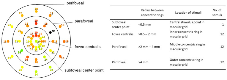 Figure 1
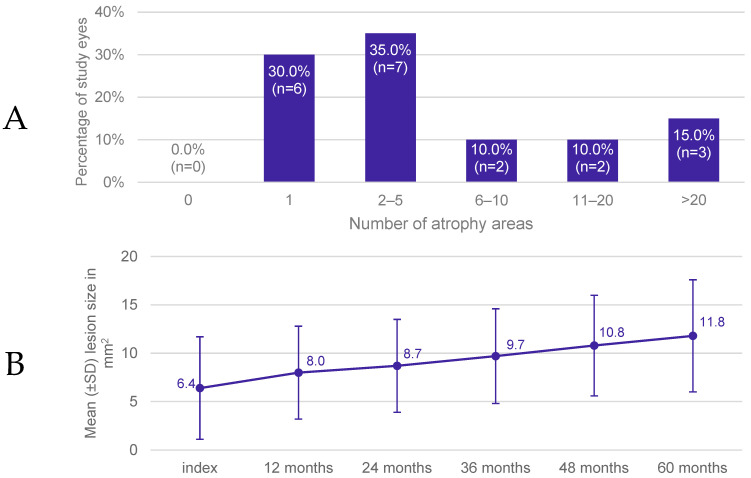 Figure 2
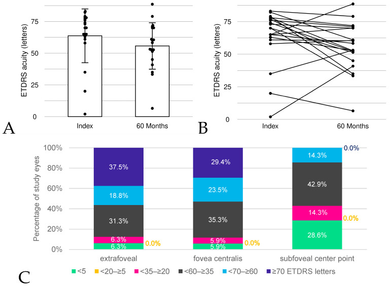 Figure 3
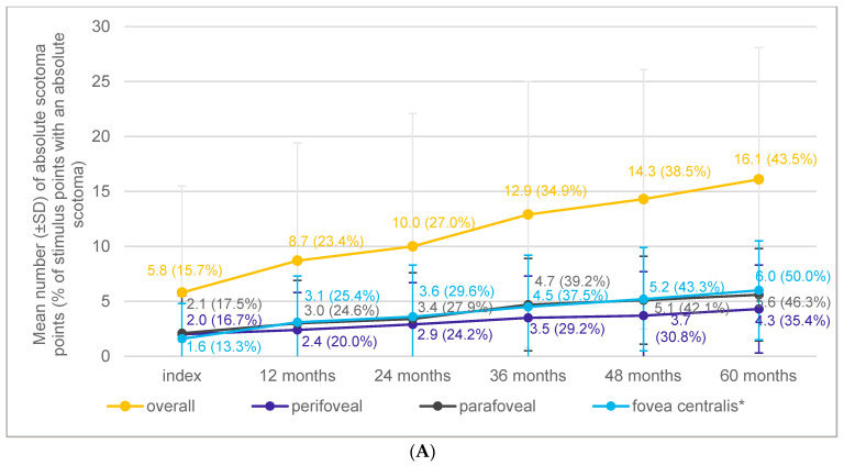 Figure 4
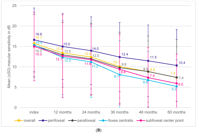 Figure 5
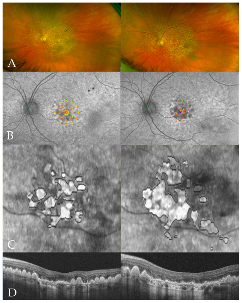 Figure 6
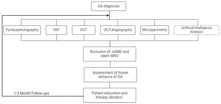 Figure 7
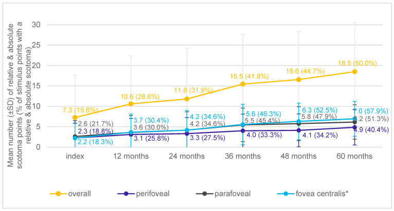 Figure 8
 Figure 9
Peer Reviews
No public reviews on file for this paper yet. If you reviewed it on a platform where reviews are public (OpenReview, ICLR, NeurIPS, ICML), you can paste yours below so the community can read it here.
Videos
No videos yet. Explain this paper in a talk, walkthrough, or lecture? Add one.
Taxonomy
TopicsRetinal Diseases and Treatments · Retinal Imaging and Analysis · Glaucoma and retinal disorders