The Usefulness of 55° Wide-Field Spectral-Domain Optical Coherence Tomography in Monitoring the Features of Peripheral Subretinal Fluid Remnants after Rhegmatogenous Retinal Detachment Surgery
Valentina Carta, Filippo Lixi, Pasquale Loiudice, Francesca Frongia, Filippo Tatti, Chiara Delpiano, Pierluca Cremonesi, Enrico Peiretti

TL;DR
This study shows that 55° wide-field OCT can effectively monitor subretinal fluid after retinal detachment surgery, especially in settings without ultra-wide-field OCT.
Contribution
Demonstrates the viability of 55° wide-field SD-OCT as an alternative for monitoring peripheral subretinal fluid in post-surgery retinal detachment patients.
Findings
55° wide-field SD-OCT detected persistent subretinal fluid in 22.5% of patients after retinal detachment surgery.
Young age was significantly associated with subretinal fluid persistence (p = 0.009).
All patients showed complete subretinal fluid resorption within 12 months of follow-up.
Abstract
Background: This study aimed to assess the effectiveness of 55° wide-field (WF) spectral-domain (SD) optical coherence tomography (OCT) for detecting peripheral subretinal fluid (SRF) after surgery for rhegmatogenous retinal detachment (RRD). Methods: In this retrospective observational study, the retinal periphery was examined to evaluate the possible presence of persistent SRF after surgery. OCT scans were acquired in infrared mode to use any peripheral vessel as a landmark for better repeatability in monitoring fluid remnants. Results: A total of 80 patients (10% with high myopia) were examined using 55° WF SD OCT after successful pars plana vitrectomy (83.8%) or scleral buckling (16.3%) for RRD. A total of 18 patients (22.5%), 16 of whom underwent pars plana vitrectomy and 2 who underwent scleral buckling, showed SRF at the OCT examination during the follow-up. Potential risk…
Genes, proteins, chemicals, diseases, species, mutations and cell lines named across the full text — each resolved to its canonical identifier and authoritative record.
Click any figure to enlarge with its caption.
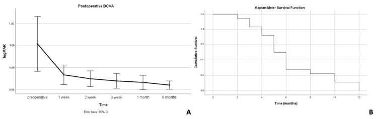 Figure 1
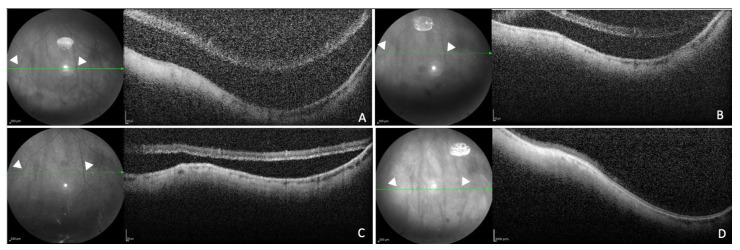 Figure 2
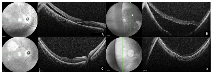 Figure 3
Peer Reviews
No public reviews on file for this paper yet. If you reviewed it on a platform where reviews are public (OpenReview, ICLR, NeurIPS, ICML), you can paste yours below so the community can read it here.
Videos
No videos yet. Explain this paper in a talk, walkthrough, or lecture? Add one.
Taxonomy
TopicsRetinal and Macular Surgery · Optical Coherence Tomography Applications · Glaucoma and retinal disorders