Central Retinal Artery Occlusion Associated with Takayasu Arteritis
Sehreen Mumtaz, Claire Wilson, Prasanna Vibhute, Eric R. Eggenberger, Florentina Berianu, Andy Abril

TL;DR
This paper discusses two cases where a rare eye condition called central retinal artery occlusion was linked to Takayasu arteritis, a rare inflammatory blood vessel disease.
Contribution
The novelty lies in presenting two specific clinical cases linking CRAO with Takayasu arteritis, highlighting a rare ocular manifestation.
Findings
Central retinal artery occlusion can be a rare but significant manifestation of Takayasu arteritis.
Only 5% of CRAO cases are arteritic, often linked to inflammatory vasculitides like Takayasu arteritis.
Ocular manifestations of Takayasu arteritis are rare and can delay diagnosis and treatment.
Abstract
Takayasu arteritis is a chronic inflammatory vasculitis with granulomatous panarteritis particularly impacting large vessels including the aorta and its branches, especially the subclavian arteries, with clinical manifestation dependent on the involved artery. Sequelae of the active disease vary, including stenosis, occlusions, or aneurysmal dilatations of the large vessels. The prevalence of Takayasu arteritis is higher in the Asian population and in Japan, but quite low in the United States, varying from 0.9–8.4 per million people. Ocular manifestations are rare and lead to a delay in diagnosis and appropriate treatment. Ocular manifestations include Takayasu retinopathy, anterior ischemic optic neuropathy (AION), retinal artery occlusion (RAO) and retinal vein occlusion (RVO). We present two cases in which central retinal artery occlusion (CRAO) was associated with Takayasu…
Genes, proteins, chemicals, diseases, species, mutations and cell lines named across the full text — each resolved to its canonical identifier and authoritative record.
Click any figure to enlarge with its caption.
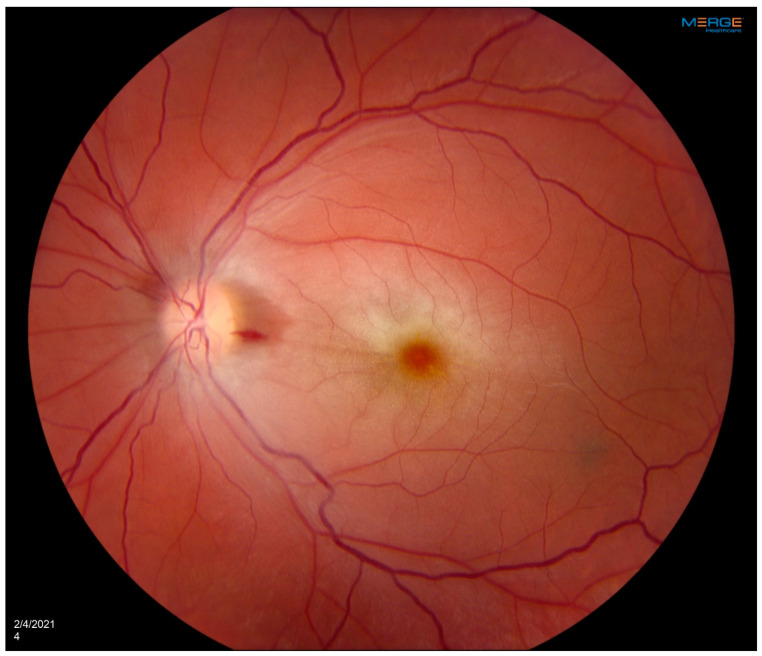 Figure 1
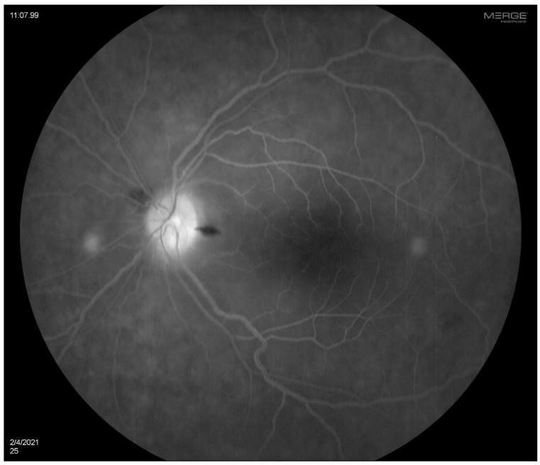 Figure 2
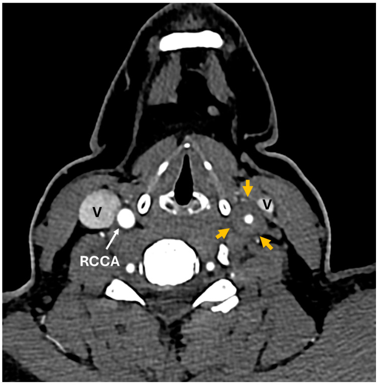 Figure 3
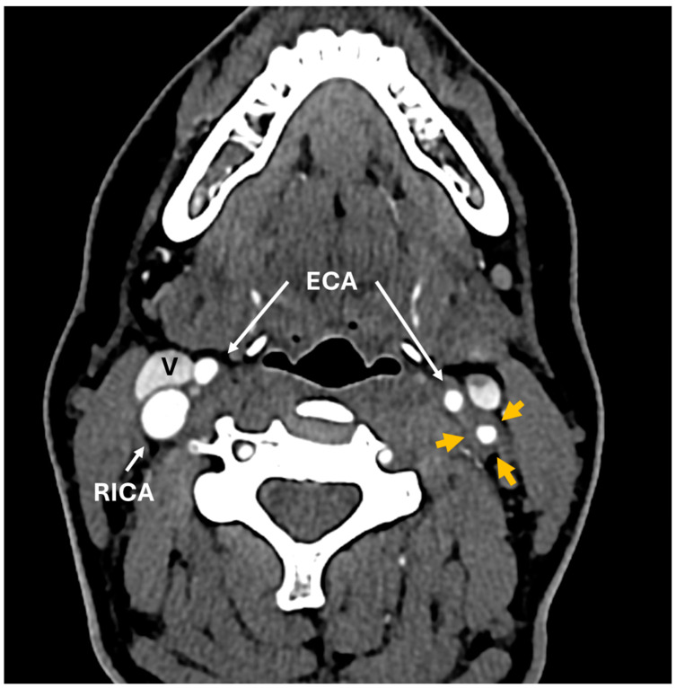 Figure 4
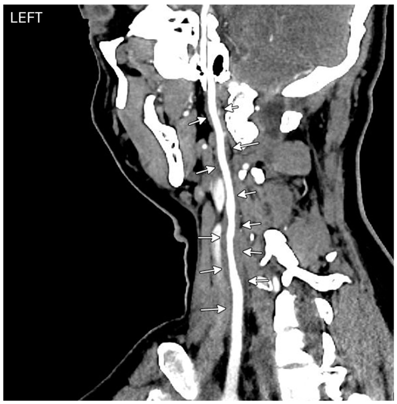 Figure 5
Peer Reviews
No public reviews on file for this paper yet. If you reviewed it on a platform where reviews are public (OpenReview, ICLR, NeurIPS, ICML), you can paste yours below so the community can read it here.
Videos
No videos yet. Explain this paper in a talk, walkthrough, or lecture? Add one.
Taxonomy
TopicsRetinal and Optic Conditions · Vasculitis and related conditions · Intraoperative Neuromonitoring and Anesthetic Effects
A 35-year-old woman with a prior history of left central retinal artery occlusion and bilateral sensorineural hearing loss, initially concerning with respect to neurosarcoidosis (never histologically proven), presented for clinical follow-up. She had a complicated disease course dating back many years, involving different specialists, and including fluctuating hearing loss partially responsive to corticosteroids necessitating cochlear implants and developed central retinal occlusion of the left eye (Figure 1 and Figure 2). This was concerning with respect to possible underlying inflammatory conditions such as neurosarcoidosis or Susac syndrome (MRI atypical sans corpus lesion), and she received IV immunoglobulin treatment. When she returned for follow-up, she had complaints of episodes of confusion, but quiescent symptoms without visual change, status post adalimumab and methotrexate addition. She sought emergency care for new transient arm numbness and confusion, and a neck CTA revealed carotid bulb circumferential thickening suggestive of vasculitis. A rind of soft tissue thickening involving the distal half of the common carotid artery, carotid bifurcation and the proximal internal and external carotid arteries was visualized. There was smooth narrowing of the encased carotid artery lumen, producing roughly 50% diameter stenosis of the distal common carotid artery. A small ulceration was noted on the left carotid bulb (Figure 3, Figure 4 and Figure 5). The patient was initiated on infliximab with IV methylprednisolone and close follow-up with neurology and vascular medicine for management of Takayasu arteritis.
A 56-year-old woman was diagnosed with Takayasu arteritis in 1991, and initially treated with glucocorticoids for several years. Initial symptoms included constitutional symptoms and left-sided carotidynia. She had markedly elevated inflammatory markers and anemia at disease onset, and later developed erythema nodosum. Imaging studies in the early phase of her illness revealed moderate stenosis of the right subclavian artery and marked thickening of the left common carotid. She was subsequently treated with methotrexate and cyclosporine, and eventually her disease went into remission. In 2014, an MR angiogram of the chest showed radiographic evidence of vessel wall edema in the aorta, and at that time, the patient was started on mycophenolate mofetil. In November 2017, the patient developed blurry vision and there was leakage on her fluorescein angiogram, which was felt to be nonspecific. She was taken off mycophenolate due to no evidence of active vasculitis in clinical or laboratory findings. She was in remission; however, she required ascending aorta hemiarch repair and aortic valve replacement (and was subsequently on warfarin) for a dilated aortic root of 61 mm with histopathology showing healed arteritis. Two years later, she developed a paracentral scotoma in the left eye and was diagnosed by outside Ophthalmology as having branch retinal artery occlusion by retinal imaging, with suspected embolic disease in the context of subtherapeutic anticoagulation.
Clinical characteristics of patients with ocular manifestations and Takayasu’s arteritis have been reported to be similar to those of patients without ocular manifestations with a mean age in the third decade of life and a majority of Asian origin. Ocular manifestations of Takayasu arteritis can be seen in a wide range (8.1–68%) of patients [1]. Apart from two case-based systematic reviews, the current literature is lacking in describing the extent of ocular involvement in Takayasu arteritis [1,2]. The current classification of Takayasu arteritis does not include visual changes, and consequently no ophthalmological examination is recommended, even after diagnosis, and ocular manifestations can go unnoticed.
A study by Pasko et al. noted ocular manifestations presenting prior to diagnosis of Takayasu arteritis in 74.6% of cases [3]. This may be explained by cases in which initial manifestations included constitutional symptoms that were non-specific. Limitation of flow in the carotid artery results in chronic ischemia and hypoperfusion retinopathy, and ocular ischemic syndrome may be of consequence [4]. It remains imperative to distinguish and categorize this ocular disorder and pursue retinal angiograms.
A study from 1994 pointed to the presence of Takayasu’s retinopathy as a statistically significant poor prognostic factor [5]. Mirouse et al. associated Takayasu retinopathy independently with mortality and complication-free survival [6]. Reportedly, 64% of CRAO patients develop new vascular risk factors after a retinal occlusive event, implying this population of patients is prone to a secondary ischemic event and needs risk factor assessment [7]. Although medical management is the recommended initial management for Takayasu arteritis, patients who have vascular complications may require surgical interventions and endovascular procedures [8]. Medical management includes ocular massage, hyperbaric oxygen therapy, and intravenous tissue-type plasminogen activator; however, these are associated with risks, including mobilization of the embolus and exacerbating macular edema. Endovascular treatments, including intra-arterial thrombolysis, have shown promising results; however, they require exceptional skill to perform [9].
Although information on ocular ischemia surgical management for Takayasu arteritis patients is limited, case series and case reports have demonstrated favorable results of balloon angioplasty and endovascular stenting in Takayasu retinopathy patients [1]. Adopting routine fundus fluorescein angiography for surveillance in patients with Takayasu arteritis may be beneficial. Newer diagnostic techniques, especially optical coherence tomography (OCT) angiography (OCTA), and novel diagnostics to assess retina perfusion must be investigated [1].
In young patients with visual loss and deficits, it is imperative to recall Takayasu arteritis as a potential suspect, even though such cases are rare, because it can translate into a meaningful difference in outcomes with proper management. Raising ophthalmological awareness of this association and increasing education regarding pursuing comprehensive diagnostic testing is crucial. Further investigation of arteritic CRAO in Takayasu arteritis patients is needed for the development and implementation of enhanced therapeutic techniques.
The reference list from the paper itself. Each links out to its DOI / PubMed record.
- 1Zeng Y. Duan J. Ge G. Zhang M. Therapeutic Management of Ocular Ischemia in Takayasu’s Arteritis: A Case-Based Systematic Review Front. Immunol.20221279127810.3389/fimmu.2021.79127835095866 PMC 8795594 · doi ↗ · pubmed ↗
- 2Sanchez-Alvarez C. Crowson C.S. Koster M.J. Warrington K.J. Prevalence of Takayasu Arteritis: A Population-based Study J. Rheumatol.20214895210.3899/jrheum.20146333589549 PMC 8456169 · doi ↗ · pubmed ↗
- 3Szydełko-Paśko U. Przeździecka-Dołyk J. NowakŁ. Małyszczak A. Misiuk-Hojło M. Ocular Manifestations of Takayasu’s Arteritis—A Case-Based Systematic Review and Meta-Analysis J. Clin. Med.202312374510.3390/jcm 1211374537297942 PMC 10253919 · doi ↗ · pubmed ↗
- 4Guclu H. Gurlu V.P. Ozal S.A. Guclu O. Central Retinal Artery Occlusion in Takayasu’s Arteritis as the First Presentation of the Disease Case Rep. Ophthalmol. Med.20162016649251310.1155/2016/649251327867673 PMC 5102719 · doi ↗ · pubmed ↗
- 5Ishikawa K. Maetani S. Long-term outcome for 120 Japanese patients with Takayasu’s disease. Clinical and statistical analyses of related prognostic factors Circulation 1994901855186010.1161/01.cir.90.4.18557923672 · doi ↗ · pubmed ↗
- 6Mirouse A. Biard L. Comarmond C. Lambert M. Mekinian A. Ferfar Y. Kahn J.-E. Benhamou Y. Chiche L. Koskas F. Overall survival and mortality risk factors in Takayasu’s arteritis: A multicenter study of 318 patients J. Autoimmun.201896353910.1016/j.jaut.2018.08.00130122419 · doi ↗ · pubmed ↗
- 7Varma D.D. Cugati S. Lee A.W. Chen C.S. A review of central retinal artery occlusion: Clinical presentation and management Eye 20132768869710.1038/eye.2013.2523470793 PMC 3682348 · doi ↗ · pubmed ↗
- 8Maz M. Chung S.A. Abril A. Langford C.A. Gorelik M. Guyatt G. Archer A.M. Conn D.L. Full K.A. Grayson P.C. 2021 American College of Rheumatology/Vasculitis Foundation Guideline for the Management of Giant Cell Arteritis and Takayasu Arteritis Arthritis Rheumatol.2021731349136510.1002/art.4177434235884 PMC 12344528 · doi ↗ · pubmed ↗