Feasibility study to unveil the potential: considerations of constrained spherical deconvolution tractography with unsedated neonatal diffusion brain MRI data
Anouk S. Verschuur, Chantal M. W. Tax, Martijn F. Boomsma, Helen L. Carlson, Gerda van Wezel-Meijler, Regan King, Alexander Leemans, Lara M. Leijser

TL;DR
This study shows that constrained spherical deconvolution tractography can reconstruct complex brain fiber structures in unsedated neonates, but results are sensitive to imaging and processing settings.
Contribution
Demonstrates the feasibility of CSD tractography for neonatal diffusion MRI and highlights the impact of processing parameters on results.
Findings
CSD tractography successfully reconstructed crossing fiber bundles in unsedated neonatal dMRI data.
Tractography results were highly sensitive to image resolution and processing settings, affecting streamline count and diffusion measures.
Diffusion measures like fractional anisotropy and mean diffusivity varied by up to 23% based on processing choices.
Abstract
The study aimed to (1) assess the feasibility constrained spherical deconvolution (CSD) tractography to reconstruct crossing fiber bundles with unsedated neonatal diffusion MRI (dMRI), and (2) demonstrate the impact of spatial and angular resolution and processing settings on tractography and derived quantitative measures. For the purpose of this study, the term-equivalent dMRIs (single-shell b800, and b2000, both 5 b0, and 45 gradient directions) of two moderate-late preterm infants (with and without motion artifacts) from a local cohort [Brain Imaging in Moderate-late Preterm infants (BIMP) study; Calgary, Canada] and one infant from the developing human connectome project with high-quality dMRI (using the b2600 shell, comprising 20 b0 and 128 gradient directions, from the multi-shell dataset) were selected. Diffusion tensor imaging (DTI) and CSD tractography were compared on b800…
Genes, proteins, chemicals, diseases, species, mutations and cell lines named across the full text — each resolved to its canonical identifier and authoritative record.
Click any figure to enlarge with its caption.
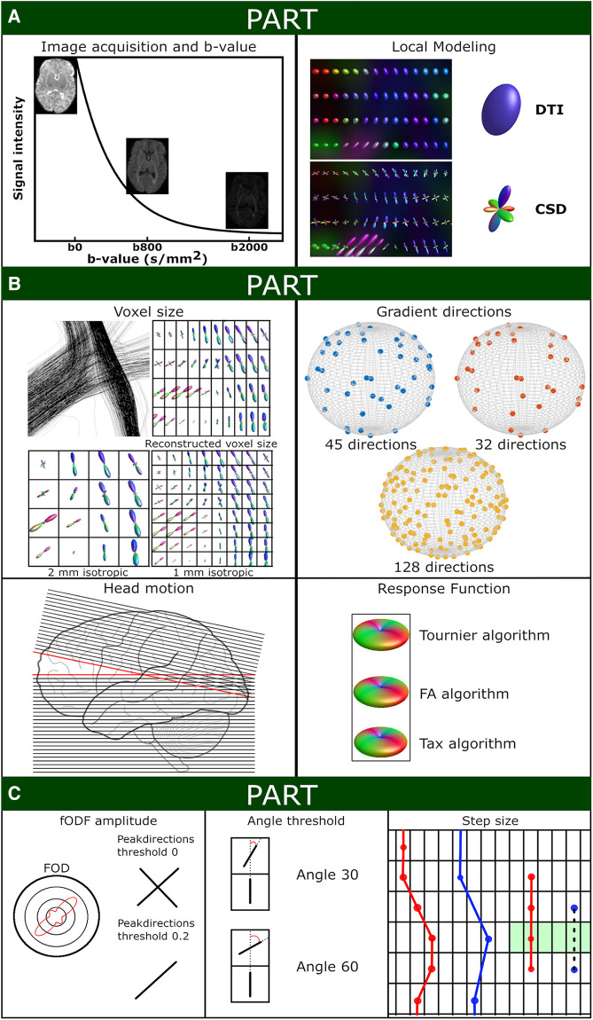 Figure 1
 Figure 2
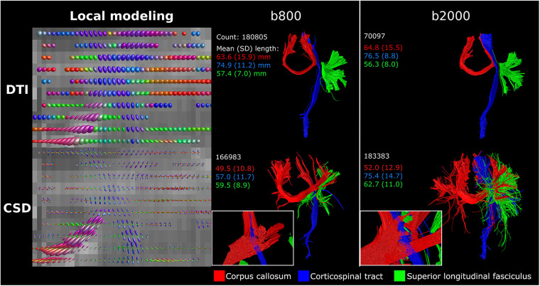 Figure 3
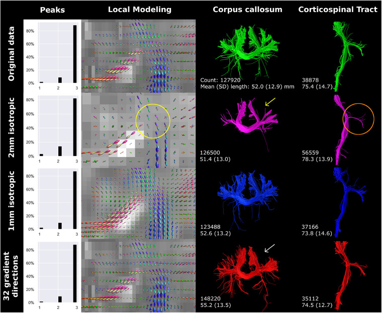 Figure 4
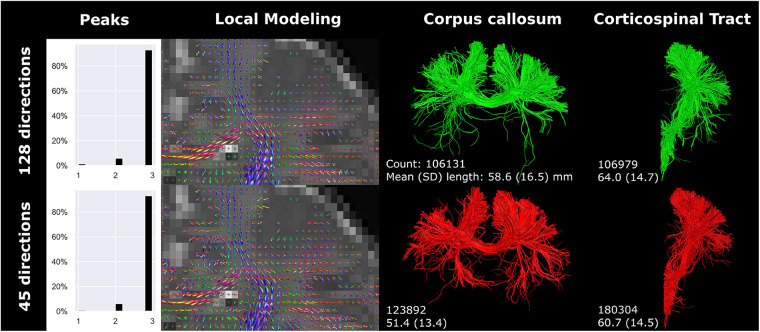 Figure 5
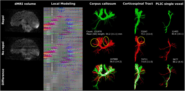 Figure 6
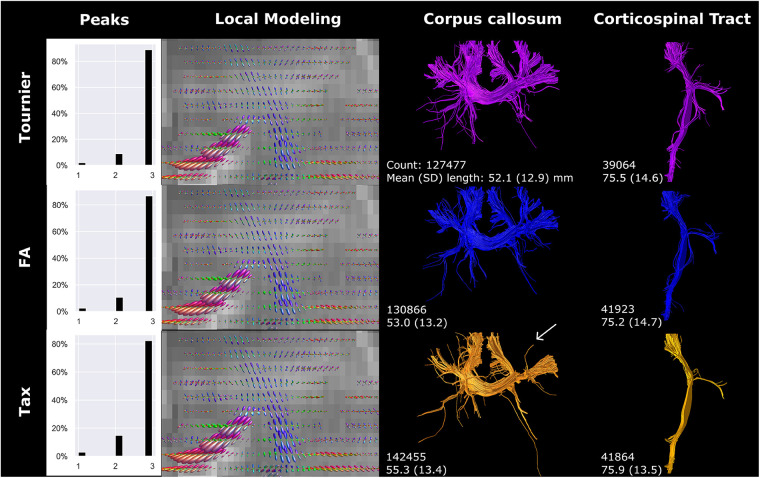 Figure 7
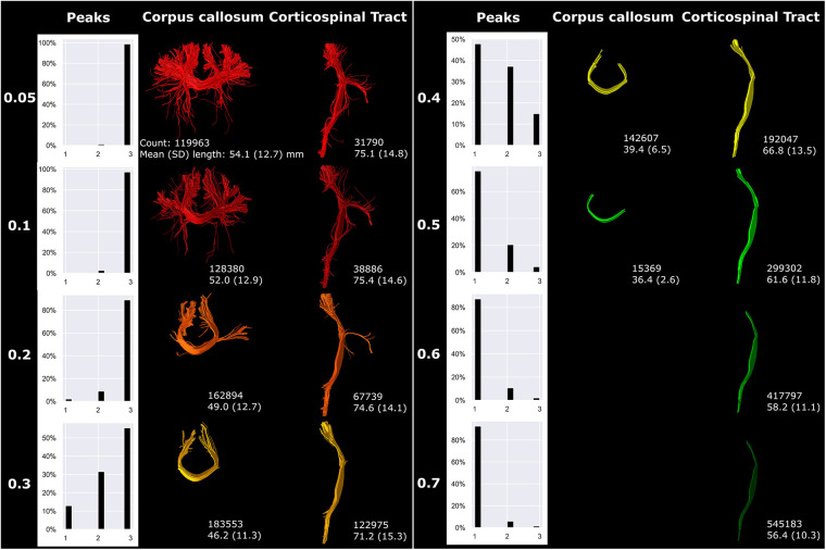 Figure 8
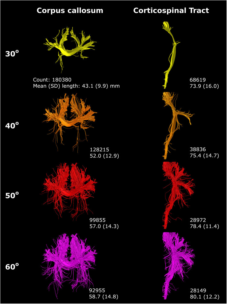 Figure 9
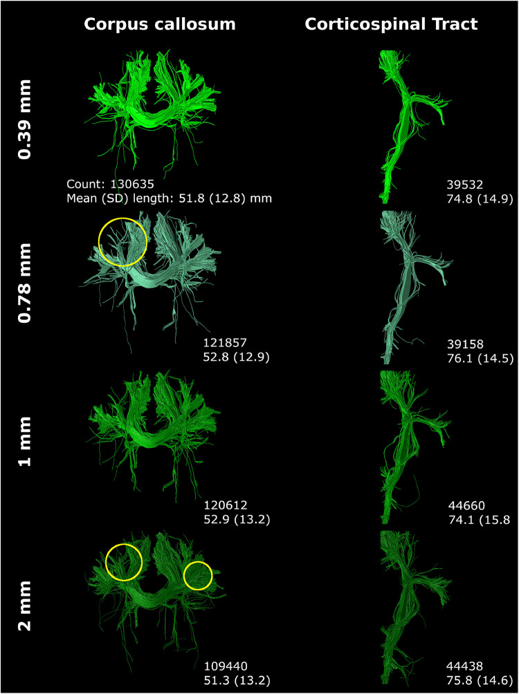 Figure 10
Peer Reviews
No public reviews on file for this paper yet. If you reviewed it on a platform where reviews are public (OpenReview, ICLR, NeurIPS, ICML), you can paste yours below so the community can read it here.
Videos
No videos yet. Explain this paper in a talk, walkthrough, or lecture? Add one.
Taxonomy
TopicsAdvanced Neuroimaging Techniques and Applications · Advanced MRI Techniques and Applications · Neonatal and fetal brain pathology