Managing a Misdiagnosed Case of Nevus Sebaceous
Kshitiz Lakhey, Namratha Puttur, Rohan Manoj, Priya Garg, Nishtha Malik

TL;DR
A young man misdiagnosed with viral warts was correctly identified with nevus sebaceous and treated with laser therapy after declining surgery.
Contribution
The case highlights the importance of accurate diagnosis and the use of histopathology to avoid mismanagement.
Findings
Nevus sebaceous was diagnosed using dermoscopic and histopathological evaluations.
Laser treatment was used as an alternative to surgical excision.
Histopathological analysis is crucial for confirming diagnosis and ruling out malignancy.
Abstract
A male patient in his early 20s presented to our outpatient clinic, having previously been misdiagnosed and unsuccessfully treated as a case of viral warts. Dermoscopic and histopathological evaluations revealed characteristic features of the nevus sebaceous. The lesion was eventually treated with an erbium-doped yttrium aluminum garnet (Er:YAG) laser after the patient declined surgical excision. Nevus sebaceous often presents with verrucous surfaces that make misdiagnosis common. A correct diagnosis is crucial due to potential neoplastic transformations. Histopathological analysis is essential for both the confirmation of disease and the exclusion of malignancy. Full-thickness surgical excision remains the preferred treatment.
Genes, proteins, chemicals, diseases, species, mutations and cell lines named across the full text — each resolved to its canonical identifier and authoritative record.
Click any figure to enlarge with its caption.
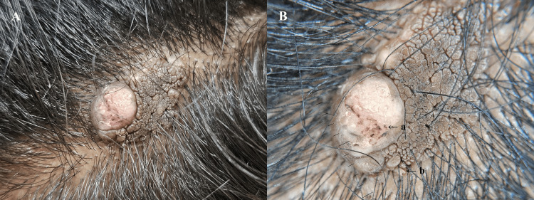 Figure 1
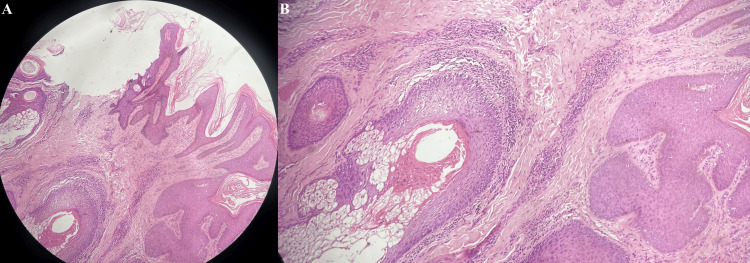 Figure 2
Peer Reviews
No public reviews on file for this paper yet. If you reviewed it on a platform where reviews are public (OpenReview, ICLR, NeurIPS, ICML), you can paste yours below so the community can read it here.
Videos
No videos yet. Explain this paper in a talk, walkthrough, or lecture? Add one.
Taxonomy
TopicsGenetic and rare skin diseases.