Decoding dose descriptors for computed tomography
Mannudeep K. Kalra, Lina Karout, Felipe de Moura Kiipper, Mônica Oliveira Bernardo

Abstract
Genes, proteins, chemicals, diseases, species, mutations and cell lines named across the full text — each resolved to its canonical identifier and authoritative record.
Click any figure to enlarge with its caption.
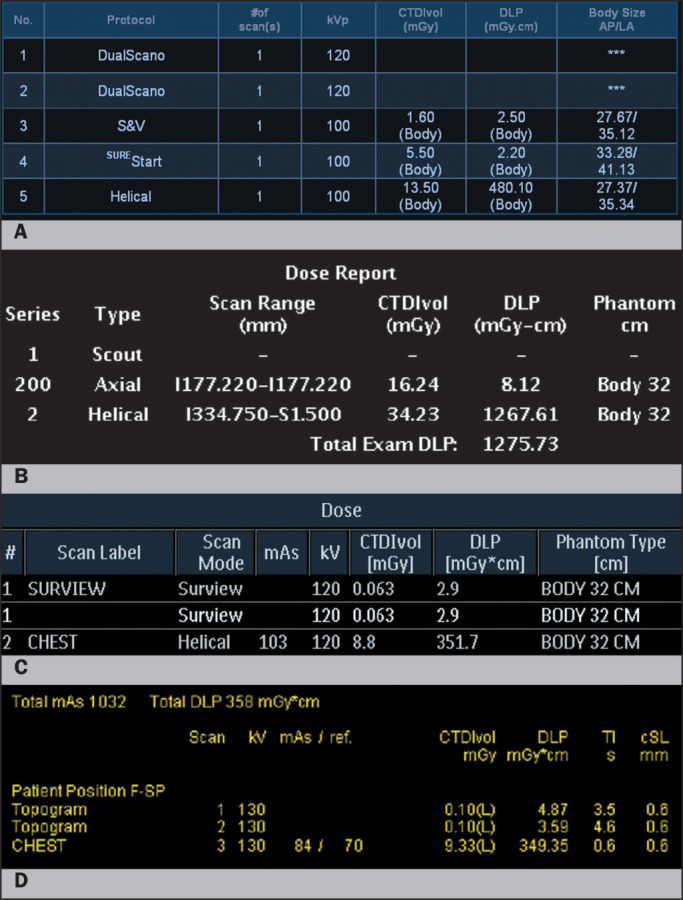 Figure 1
| Body region | Clinical indication | Descriptor | |||
|---|---|---|---|---|---|
| CTDIvol (mGy) | DLP (mGy.cm) | ||||
| ADC| | DRLCI | ADC| | DRLCI | ||
| Head | Head trauma | 26 | 37 | 601 | 769 |
| Headache | 29 | 45 | 616 | 955 | |
| Stroke | 30 | 53 | 617 | 998 | |
| Head CTA | 19 | 26 | 633 | 1,181 | |
| Paranasal sinus | Sinusitis | 15 | 19 | 228 | 353 |
| Cervical spine | Trauma | 16 | 23 | 394 | 547 |
| Chest | COVID-19 | 8 | 12 | 320 | 454 |
| Lung cancer | 6 | 10 | 249 | 419 | |
| Pneumonia | 8 | 10 | 285 | 379 | |
| Pulmonary embolism | 11 | 15 | 376 | 582 | |
| Abdomen | Appendicitis | 9 | 12 | 443 | 632 |
| Kidney stones | 10 | 13 | 535 | 717 | |
| Chest and abdomen | Cancer | 8 | 13 | 628 | 1,345 |
| Examination | Age (years) | CTDIvol (mGy) | DLP (mGy.cm) | ||
|---|---|---|---|---|---|
| DRL (AD) | Range | DRL (AD) | Range | ||
| Unenhanced | 0-< 1 | 27 (20) | 3-113 | 456 (302) | 35-1508 |
| head CT | l-< 2 | 30 (20) | 5-113 | 535 (356) | 104-2528 |
| 2-< 6 | 44 (29) | 8-70 | 813 (527) | 120-1650 | |
| 6-18 | 52 (35) | 9-77 | 949 (625) | 82-3023 | |
| Unenhanced | 0-< 1 | 4(2) | 0.19-14.0 | 81 (39) | 3-230 |
| chest CT | l-<5 | 4(2) | 0.2-24.0 | 96 (49) | 4-810 |
| 5-< 10 | 5(4) | 0.3-28.0 | 176(120) | 8-743 | |
| 10-< 15 | 8(5) | 0.4-28.0 | 294 (172) | 9-1828 | |
| 15-18 | 11 (6) | 0.9-22.0 | 425 (276) | 19-1168 | |
| Contrast- | 0-< 1 | 5(2) | 0.2-13.0 | 83 (42) | 3-277 |
| enhanced | l-<5 | 5(3) | 0.2-39.0 | 110 (62) | 6-916 |
| chest CT | 5-< 10 | 6(4) | 0.3-28.0 | 167 (107) | 9-785 |
| 10-< 15 | 9(6) | 0.5-47.0 | 293 (202) | 9-2077 | |
| 15-18 | 12 (8) | 0.3-35.0 | 412 (305) | 13-1846 | |
| Contrast- | 0-< 1 | 3(2) | 0.2-42.0 | 130 (46) | 3-386 |
| enhanced | l-<5 | 4(3) | 2-29 | 160 (103) | 9-794 |
| abdomen-pelvis CT | 5-< 10 | 7(4) | 0.6-46.0 | 260 (163) | 17-1298 |
| 10-< 15 | 12 (7) | 1-80 | 418 (292) | 39-3027 | |
| 15-18 | 13 (9) | 2.5-78.0 | 529 (414) | 97-2028 | |
Peer Reviews
No public reviews on file for this paper yet. If you reviewed it on a platform where reviews are public (OpenReview, ICLR, NeurIPS, ICML), you can paste yours below so the community can read it here.
Videos
No videos yet. Explain this paper in a talk, walkthrough, or lecture? Add one.
Taxonomy
TopicsRadiation Dose and Imaging · Medical Imaging Techniques and Applications · Advanced X-ray and CT Imaging
INTRODUCTION
The Latin Safe initiative and the Massachusetts General Hospital Webster Center for Quality and Safety present a series of News in Radiology articles encompassing different aspects of radiation dose optimization in diagnostic imaging. Each article will present practical aspects that can help radiologists, technologists, and medical physicists understand and apply those concepts to improve patient safety and quality. This first article explains radiation dose descriptors for computed tomography (CT).
RADIATION DOSE DESCRIPTORS
Regulations require all CT scanners to display scanner output values^(1)^, including the volume CT dose index (CTDIvol, in mGy) and dose length product (DLP, in mGy.cm)-Figure 1. The CTDIvol represents the average radiation dose estimated by using polymethyl methacrylate phantoms of either 16 cm or 32 cm (typically for head and body CT, respectively). The DLP, defined as the total dose over the entire length of a scan, is the product of the CTDIvol and scan length.
Figure 1. Dose information pages from four major CT vendors (A: Canon; B: GE; C: Philips; D: Siemens). Despite their differences, each vendor provides series-specific CTDIvol and DLP, as well as the corresponding phantom size, which enables users to compare radiation doses across different CT protocols and scanners. AP, anterior-posterior; LA, lateral; TI, (tube rotation) time; cSL, slice collimation; F-SP, feet first-supine.
The size-specific dose estimates derived from multiplying CTDIvol by a conversion factor based on patient size (measured diameters) compensate for the variation in patient sizes^(2)^. Diagnostic reference levels (DRLs) represent 75th-percentile radiation dose indices (CTDIvol and DLP) for a specific body region, examination type, or clinical indication, obtained from a survey of radiation doses at the local, regional, national, or international level^(3)^. The effective dose, a term coined by the International Commission of Radiation Protection, describes the relative whole-body radiation dose (in mSv), which is estimated by summing the individual organ doses or, more crudely, by multiplying the DLP by the conversion coefficients specific to the age of the patient and the body segment being imaged. Calculation of the effective dose allows radiation risks to be evaluated at the population level.
Limitations
The CTDIvol and DLP represent absorbed doses in phantoms or the scanner output dose, rather than patient doses. These doses should not be used to estimate radiation risks associated with CT scanning. The DRLs should not be used for optimizing radiation doses on a per-patient basis, because they do not account for variations in patient size or scanner-specific attributes.
Practical applications
Despite their limitations, CT radiation dose descriptors are powerful tools in radiation dose optimization. Because these descriptors are measured in a standardized manner, they allow dose comparison across all scanners and CT protocols. Given that they are displayed during the planning of the examination and before the actual scanning, users can modify scan factors to adjust the doses prior to scanning. In addition, the dose descriptors can be obtained either from a dose information page or from a structured Digital Imaging and Communications in Medicine report, either manually or automatically with radiation dose monitoring software. Dose monitoring allows institutions to compare their doses with those of other institutions or against internal target dose values.
Radiation doses are linked to image quality, and the specific clinical indication or motive should dictate the quality of the image. Therefore, the monitoring of radiation doses must involve documentation and analysis of the clinical indications or reasons for scanning^(4,5)^. With the technological revolution proceeding at an exponential pace, most scanners can automatically adapt radiation doses and maintain quality for different body parts and patient sizes. However, such automation fails if users do not make manual adjustments based on the clinical indications. It is imperative that all interpreting physicians and CT technologists know and understand the typical local values for CTDIvol and DLP at their institutions, as well as how those values compare with the regional, national, and international DRLs (Tables 1 and 2).
Table 1: Clinical indication-based DRLs in Brazil from a multicenter effort led by the Brazilian College of Radiology and Diagnostic Imaging. Although the DRLs for head CT are well below those employed in the United States and Europe, the DRLs for chest and abdomen CT suggest a need for protocol optimization and radiation dose reduction.
Table 2: Summary of radiation doses for pediatric CT examinations in Latin America.
CONCLUSION
Dose descriptors for CT are a powerful ally in the quest for radiation dose optimization. Radiologists and technologists should understand the strengths and weaknesses of these descriptors.
The reference list from the paper itself. Each links out to its DOI / PubMed record.
- 1Kalra MK Sodickson AD Mayo-Smith WW. CT radiation: key concepts for gentle and wise use Radiographics 201535170617212646618010.1148/rg.2015150118 · doi ↗ · pubmed ↗
- 2AAPM Size-specific dose estimates (SSDE) in pediatric and adult body CT examinations. Report of AAPM Task Group 204College Park, MD American Association of Physicists in Medicine 2011
- 3Cadavid L Karout L Kalra MK Setting up regional diagnostic reference levels for pediatric computed tomography in Latin America: preliminary results, challenges and the work ahead Pediatr Radiol 2024544574673722746610.1007/s 00247-023-05676-9 · doi ↗ · pubmed ↗
- 4Bernardo M Homayounieh F Cuter MCR Chest CT usage in COVID-19 pneumonia: multicenter study on radiation doses and diagnostic quality in Brazil Radiat Prot Dosimetry 20211971351453487569210.1093/rpd/ncab 171PMC 8903326 · doi ↗ · pubmed ↗
- 5Rastogi S Singh R Borse R Use of multiphase CT protocols in 18 countries: appropriateness and radiation doses Can Assoc Radiol J 2021723813873206300910.1177/0846537119888390 · doi ↗ · pubmed ↗