CT and MRI Findings of Renal Angiomyolipoma With Lung Metastasis: A Case Report and Literature Review
Zhiqiang He, Jing Wu, Xiao ming Fu, Xiao ran Li, Hai Xu, Yu‐Chen Chen

TL;DR
This case report presents a rare instance of kidney tumor with lung spread, using both CT and MRI for diagnosis.
Contribution
First reported case combining CT and MRI for diagnosing renal angiomyolipoma with lung metastasis.
Findings
Renal epithelioid angiomyolipoma with lung metastasis was diagnosed using CT and MRI.
Combining CT and MRI provides a novel approach for diagnosing this rare condition.
Abstract
Renal angiomyolipoma has two histological variants: classical and epithelioid. Epithelioid angiomyolipoma is considered as a potential malignant tumor, often leading to recurrence and metastasis, with rapid progression in most of the cases. The lung is one of the most commonly reported sites of metastasis, and pulmonary metastasis of renal angiomyolipoma is usually diagnostic by computed tomography (CT) scans. Here, we report for the first time renal angiomyolipoma with lung metastasis by combining CT and magnetic resonance imaging (MRI). We report for the first time renal epithelioid angiomyolipoma with lung metastasis by combining CT and magnetic resonance imaging (MRI).
Genes, proteins, chemicals, diseases, species, mutations and cell lines named across the full text — each resolved to its canonical identifier and authoritative record.
Click any figure to enlarge with its caption.
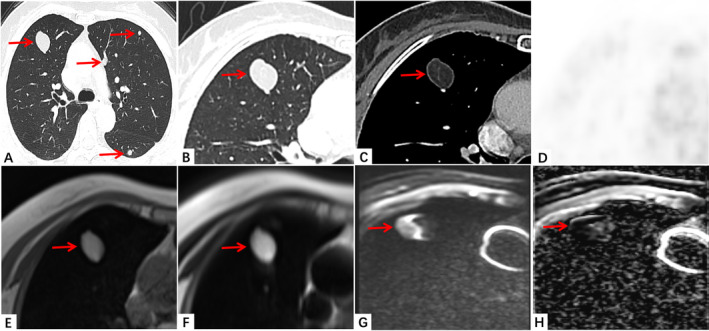 Figure 1
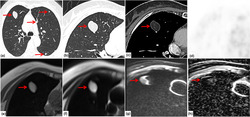 Figure 2
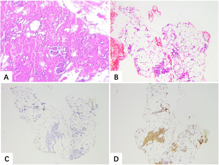 Figure 3
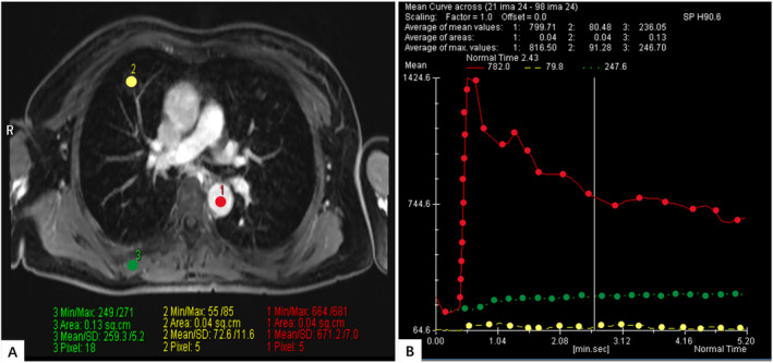 Figure 4
Peer Reviews
No public reviews on file for this paper yet. If you reviewed it on a platform where reviews are public (OpenReview, ICLR, NeurIPS, ICML), you can paste yours below so the community can read it here.
Videos
No videos yet. Explain this paper in a talk, walkthrough, or lecture? Add one.
Taxonomy
TopicsTuberous Sclerosis Complex Research · Renal cell carcinoma treatment · Renal and related cancers