Pulmonary Cystic Echinococcosis: A Ruptured and Infected Cyst Presenting As Pyopneumothorax
Tithi S, Pankaj Wagh

TL;DR
A 62-year-old woman with a ruptured lung cyst caused by Echinococcus was treated with drainage and medication.
Contribution
The case highlights a rare presentation of pulmonary cystic echinococcosis as pyopneumothorax.
Findings
Contrast-enhanced CT and chest X-ray confirmed a ruptured hydatid cyst with pyopneumothorax.
Echinococcus antibody IgG test supported the diagnosis.
Conservative treatment with drainage and albendazole was effective.
Abstract
In this report, a case of 62-year-old female is described who came to the hospital with chief complaints of breathlessness and productive cough with salty whitish expectoration, which she had for two months, along with fever and right-sided chest pain, for three days. The case was identified as a ruptured pulmonary hydatid cyst with pyopneumothorax using contrast-enhanced computed tomography and chest X-ray. This was further supported by the Echinococcus antibody IgG test. Right thoracostomy, the placement of an intercoastal drain, and four days of continuous aspiration of 750 ml of serous fluid were used for managing the case. Following this, oral albendazole was used as a conservative measure.
Genes, proteins, chemicals, diseases, species, mutations and cell lines named across the full text — each resolved to its canonical identifier and authoritative record.
Click any figure to enlarge with its caption.
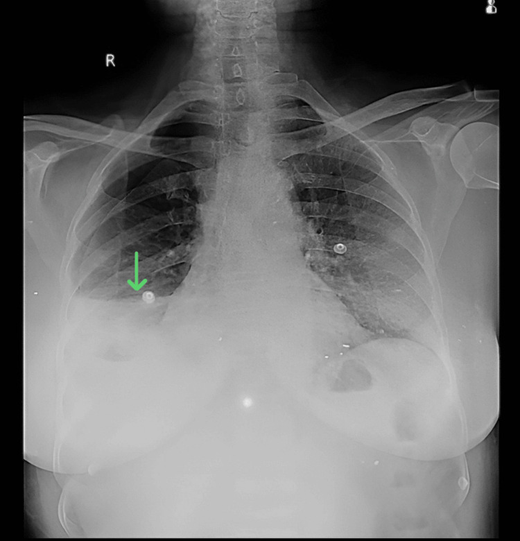 Figure 1
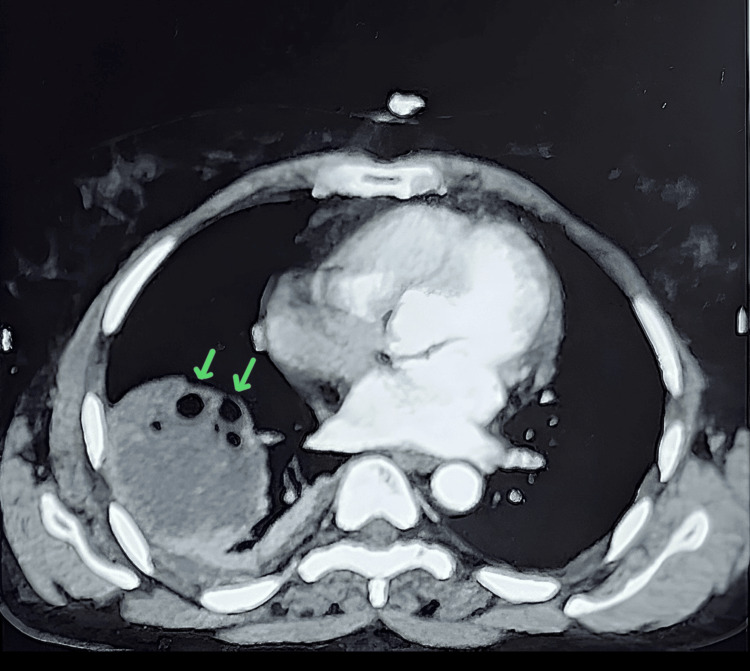 Figure 2
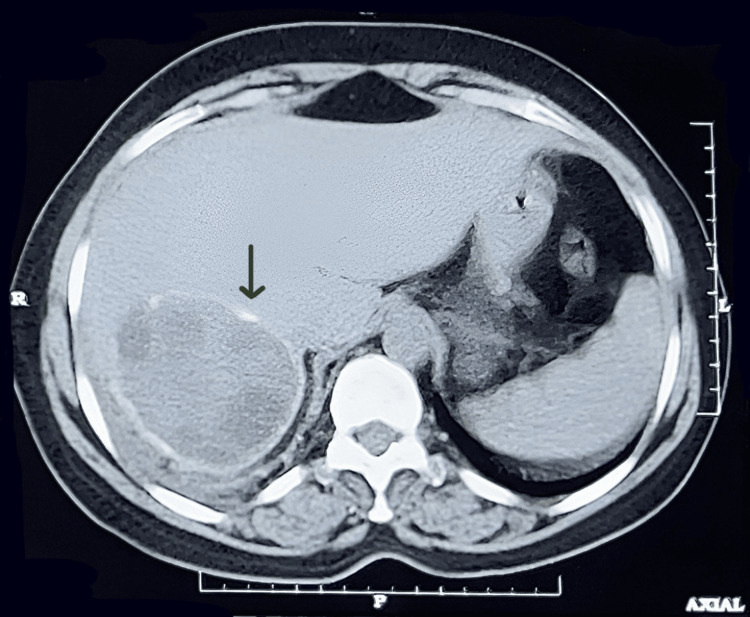 Figure 3
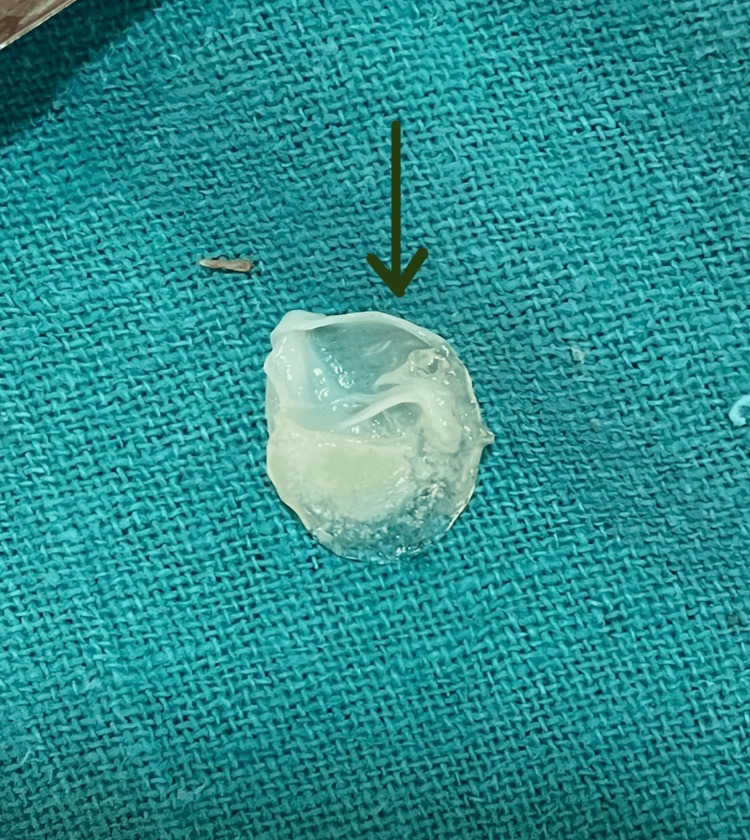 Figure 4
Peer Reviews
No public reviews on file for this paper yet. If you reviewed it on a platform where reviews are public (OpenReview, ICLR, NeurIPS, ICML), you can paste yours below so the community can read it here.
Videos
No videos yet. Explain this paper in a talk, walkthrough, or lecture? Add one.
Taxonomy
TopicsFrench Literature and Poetry