Case Report: Urethral Diverticulum after Distal Hypospadias Repair: An Uncommon Complication
Ammar Kheyami, Abdulla AlMuharraqi, Mahmood Abbas, A.K Singhal, Mohamed Zouari, Abdulla AlMuharraqi, Johannes Wirmer, Abdulla AlMuharraqi

TL;DR
A 2-year-old child developed a rare complication called urethral diverticulum after hypospadias repair surgery, which was successfully treated with surgery and resolved completely.
Contribution
This case report highlights urethral diverticulum as a rare but important complication of hypospadias repair in children.
Findings
The child presented with voiding dysfunction and was diagnosed with a urethral diverticulum via ultrasound and cystourethroscopy.
Surgical intervention resolved the symptoms, with no recurrence observed after 3 months of follow-up.
The report emphasizes the importance of early detection and awareness of this complication among parents and physicians.
Abstract
A urethral diverticulum can be defined as a pocket that forms from the lining of the urethra and protrudes into the surrounding tissue, a condition which causes voiding dysfunction and may result as a rare complication of hypospadias repair surgery. We report the case of a 2-year-old child who presented to us in 2019 complaining of a thin forceful stream, ballooning of the ventral aspect of the penis while voiding, and post-void dribbling. He has a history of undergoing a tabularised incised plate urethroplasty for distal penile hypospadias at 18-months-old. Ultrasound showed increased post-void residual volume and cystourethroscopy confirmed a urethral diverticulum extending from the subcorona to the base of the penis. The patient underwent partial excision of diverticulum, urethroplasty, and meatoplasty. He was followed-up 3 months later with complete resolution of his symptoms and a…
Genes, proteins, chemicals, diseases, species, mutations and cell lines named across the full text — each resolved to its canonical identifier and authoritative record.
Click any figure to enlarge with its caption.
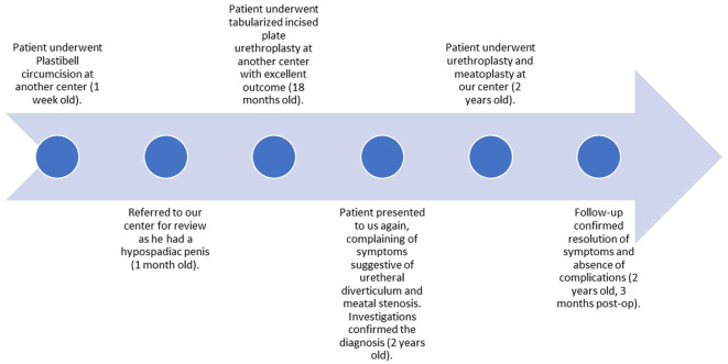 Figure 1
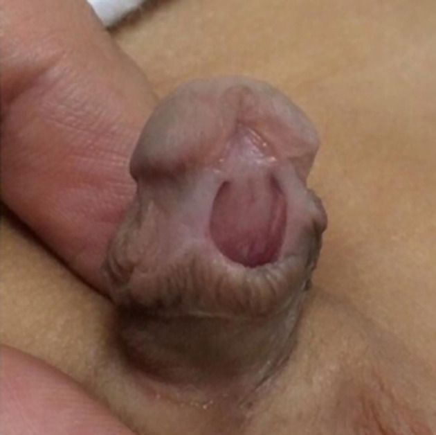 Figure 2
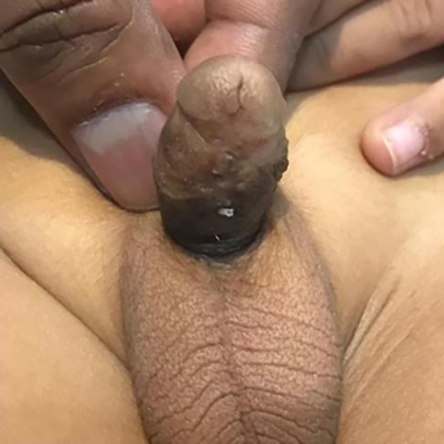 Figure 3
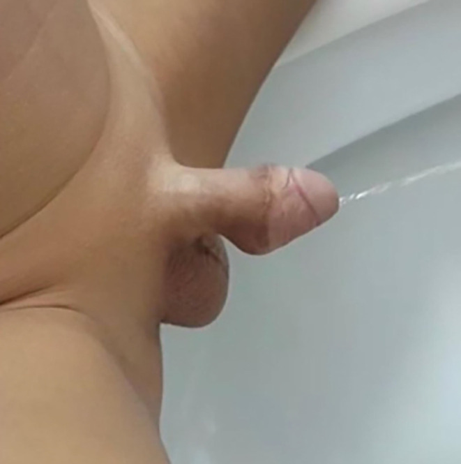 Figure 4
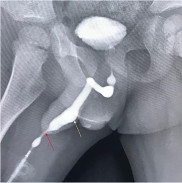 Figure 5
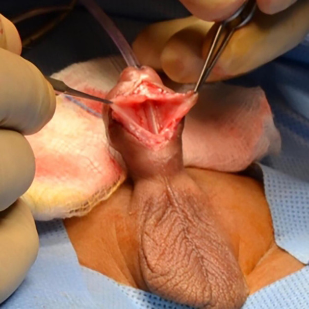 Figure 6
Peer Reviews
No public reviews on file for this paper yet. If you reviewed it on a platform where reviews are public (OpenReview, ICLR, NeurIPS, ICML), you can paste yours below so the community can read it here.
Videos
No videos yet. Explain this paper in a talk, walkthrough, or lecture? Add one.
Taxonomy
TopicsUrological Disorders and Treatments · Urinary and Genital Oncology Studies · Urologic and reproductive health conditions
Introduction
Urethral diverticulum - an out-pouching of the urethra into surrounding tissues - is an important but uncommon complication of hypospadias repair. ^ 1 ^ Studies suggest that it comprises 0.3% of postoperative complications and is mainly associated with proximal hypospadias. ^ 2 ^ ^,^ ^ 3 ^ Factors predisposing to its formation include proximal defect, oversized neourethra, poorly supporting tissue covering urethroplasty, and distal urethral stricture. ^ 2 ^ ^,^ ^ 3 ^ We present a case of postoperative urethral diverticulum formation after distal hypospadias repair and discuss possible treatment options.
Case report
The child was one-month-old ( Figure 1) when referred to our centre in 2019 for review after undergoing Plastibell circumcision. On examination, he was circumcised with a hypospadiac meatus located at distal penile position ( Figure 2).
A timeline of the patient’s presentations and the procedures he underwent.
Ventral view of the penis (excessively circumcised with a distal penile patulous meatus).
At 18 months of age, he underwent a tabularised incised plate urethroplasty at an external paediatric urology centre with an excellent outcome ( Figure 3).
Ventral view of the penis after tabularised incised plate urethroplasty.
One year later, he presented with typical signs and symptoms of a urethral diverticulum and meatal stenosis; thin forceful stream, ballooning of the ventral aspect of the penis during voiding, significant post-void dribbling, and need for manual emptying of the diverticulum ( Figure 4).
Penis with urethral diverticulum.
Ultrasound showed diffuse bladder wall thickening with significant post-void residual volume. Ascending urethrogram confirmed the diagnosis of meatal stenosis with proximal diverticulum ( Figure 5). Patient underwent cystourethroscopy which confirmed a diverticulum extending from the subcorona to the base of the penis. The diverticulum was opened through a ventral midline incision. A midline strip of 16 mm width of mucosa was preserved, and lateral mucosa was excised preserving the underlying dartos tissue ( Figure 6).
Ascending urethrogram showing meatal narrowing (red arrow) and a proximal urethral diverticulum (yellow arrow).
Ventral view of the penis during standard meatoplasty.The meatus was opened wider and stitches were taken at the margins.
The urethroplasty was closed in two layers and was supported by double-breasted Dartos fascia. At the end of the procedure, standard meatoplasty was performed for meatal stenosis. Three months following repair, the child had a normal thick continuous stream with no post-void dribbling or ballooning of the urethra.
Discussion
Urethral diverticulum is an uncommon complication of hypospadias surgery which has several risk factors that could influence its incidence such as a large neourethra, inadequate supporting tissue post-urethroplasty, and a distal urethral stricture. ^ 2 ^ ^,^ ^ 3 ^ It can be diagnosed via clinical evaluation and confirmed with cystourethrography and cystoscopy. Partial excision of diverticulum and urethroplasty is an acceptable form of treatment for such cases, as the final aim is to restore a urethra with normal diameter and pressure that is also well supported with enough tissue and no distal obstruction. ^ 2 ^ ^,^ ^ 4 ^ In the presented case, the diagnosis of hypospadias was missed at birth and a circumcision was carried out though circumcision is contraindicated in these cases. This certainly requires more awareness since the repair of hypospadias in uncircumcised children is technically easier and has a better outcome. Additionally in this case, excessive circumcision resulted in a very wide meatus along with a severe deficiency in the ventral skin and the underlying supportive tissue which possibly complicated the repair and played an important role in the formation of the diverticulum. Meatal stenosis is another factor that predisposed to diverticulum formation in this child, and it was most likely a post-surgical complication as it can occur after any hypospadias surgery. Therefore, regular follow-up after hypospadias repair is of vital importance for early diagnosis and management of meatal stenosis.
Conclusion
Meatal stenosis and urethral diverticulum are amongst the possible complications of hypospadias repair. The repair of urethral diverticula is challenging, and the outcome depends on the creation of a well-supported and normal-sized urethra with no distal obstruction. Furthermore, parents should be educated about the signs and symptoms of meatal stenosis and the need for strict and regular follow-up to diagnose and manage any complication as early as possible. It is important to carefully examine the child for hypospadias before circumcision as it is contraindicated.
Consent
We discussed and explained to the patient’s father about the writing and publication of this case report. He had the opportunity to ask questions about everything related to this process, and understood that no identifying information related to the patient will be shared; Written informed consent for the publication of patient details and images was obtained from the patient’s father.
The reference list from the paper itself. Each links out to its DOI / PubMed record.
- 1Crescenze IM Goldman HB : Female Urethral Diverticulum: Current Diagnosis and Management. Curr. Urol. Rep. 2015;16:71. 10.1007/s 11934-015-0540-8 26267225 · doi ↗ · pubmed ↗
- 2Snodgrass W : Tubularized incised plate hypospadias repair: indications, technique, and complications. Urology. 1999;54:6–11. 10.1016/S 0090-4295(99)00144-2 10414718 · doi ↗ · pubmed ↗
- 3Snyder CL Evangelidis A Snyder RP : Management of urethral diverticulum complicating hypospadias repair. J. Pediatr. Urol. 2005;1:81–83. 10.1016/j.jpurol.2004.10.001 18947541 · doi ↗ · pubmed ↗
- 4Bote SM Gite VA Siddiqui AK : Acquired male urethral diverticulum: Report of two cases. Arch. Int. Surg. 2016;6:54–56. 10.4103/2278-9596.187204 · doi ↗
- 5: 'Pseudospongioplasty' in the repair of a urethral diverticulum. BJU Int .2004;94(1) : 10.1111/j.1464-410X.2003.04913.x 126-30 10.1111/j.1464-410X.2003.04913.x 15217446 · doi ↗ · pubmed ↗
- 6: Complications of hypospadias surgery. Clin Plast Surg .1988;15(3) :371-9 3134152 · pubmed ↗
- 7: Management of urethral diverticulum complicating hypospadias repair. J Pediatr Urol .2005;1(2) : 10.1016/j.jpurol.2004.10.001 81-3 10.1016/j.jpurol.2004.10.001 18947541 · doi ↗ · pubmed ↗
- 8: Surgical Correction of Post-hypospadias Repair Urethral Diverticulum and Stricture using Rotational Flap. Korean Journal of Urology .2006;47(11) : 10.4111/kju.2006.47.11.1255 10.4111/kju.2006.47.11.1255 · doi ↗