Author Correction: Using organ-on-a-chip technology to study haemorrhagic activities of snake venoms on endothelial tubules
Mátyás A. Bittenbinder, Flavio Bonanini, Dorota Kurek, Paul Vulto, Jeroen Kool, Freek J. Vonk

Abstract
Genes, proteins, chemicals, diseases, species, mutations and cell lines named across the full text — each resolved to its canonical identifier and authoritative record.
Click any figure to enlarge with its caption.
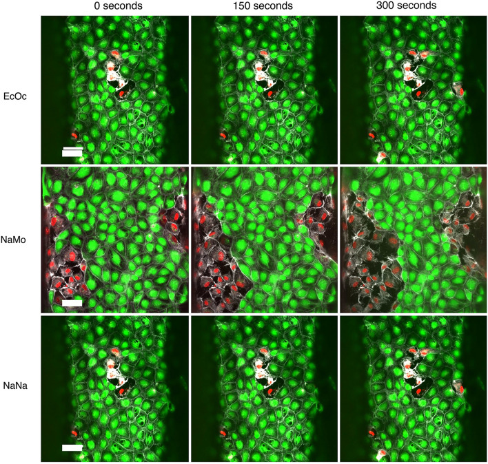 Figure 6
Peer Reviews
No public reviews on file for this paper yet. If you reviewed it on a platform where reviews are public (OpenReview, ICLR, NeurIPS, ICML), you can paste yours below so the community can read it here.
Videos
No videos yet. Explain this paper in a talk, walkthrough, or lecture? Add one.
Taxonomy
TopicsVenomous Animal Envenomation and Studies
Correction to: Scientific Reports, 10.1038/s41598-024-60282-5, published online 4 June 2024
The original version of this Article contained an error in Figure 6, where the pictures of the cells in the top row (EcOc) and the bottom row (NaNa) were duplicated. The original Figure 6 and accompanying legend appear below.Figure 6. Timelapse of high venom dose exposure on endothelial tubules Immunofluorescent microscopy images show the difference in morphology of the endothelial vessels after 0, 150, and 300 s of exposure to 100 μg/mL of snake venom compared to the control. PI is shown in red, Calcein-AM is shown in green, and live-actin is shown in white. The scale bar represents 50 μm.
The original Article has been corrected.