Rehabilitation Protocol for Lunate Fracture in a Clinical Case Report
Chitwan S Agrawal, Mitushi Deshmukh, Sakshi Padmawar

TL;DR
This paper presents a rehabilitation protocol for a patient with a lunate fracture, showing improved function and quality of life after surgery and physiotherapy.
Contribution
The paper introduces a successful rehabilitation approach for lunate fracture patients following proximal row carpectomy.
Findings
Post-operative physiotherapy significantly improved the patient's functional activities and quality of life.
Proximal row carpectomy followed by rehabilitation can be effective for lunate fracture treatment.
Natural healing is possible with decompression osteotomies to prevent lunate collapse.
Abstract
A carpal injury called a lunate fracture can cause severe carpal instability if treatment is not received. After the scaphoid, triquetrum, and trapezium, the lunate is the fourth most frequently fractured carpal bone. Due to lunate fracture, the functional prognosis is uncertain, and conservative treatment frequently results in surgery. Lunate fracture may be caused by anatomical features such as ulnar and radial variation, although necrosis is not commonly one of them. Vascularization in the lunate is unstable and dependent on the capsular arterioles. High shear stresses are frequently applied to the lunate, which rubs against the triangular fibrocartilage complex and the radius. This could lead to a fracture. Intraosseous compartment syndrome is most likely caused by the inflammatory condition Kienböck's disease. In order to protect the lunate against shear and compression loads,…
Genes, proteins, chemicals, diseases, species, mutations and cell lines named across the full text — each resolved to its canonical identifier and authoritative record.
Click any figure to enlarge with its caption.
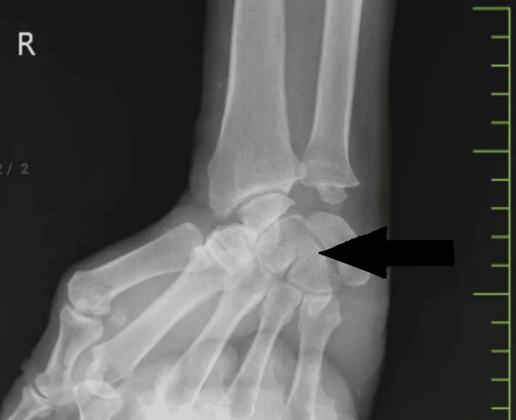 Figure 1
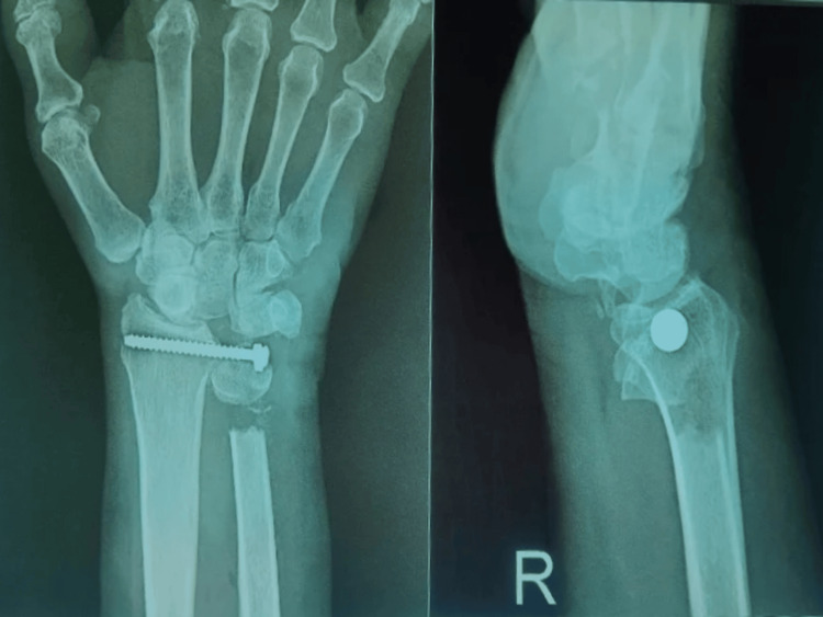 Figure 2
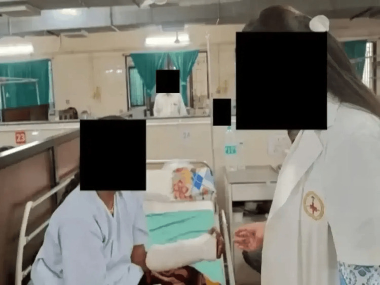 Figure 3
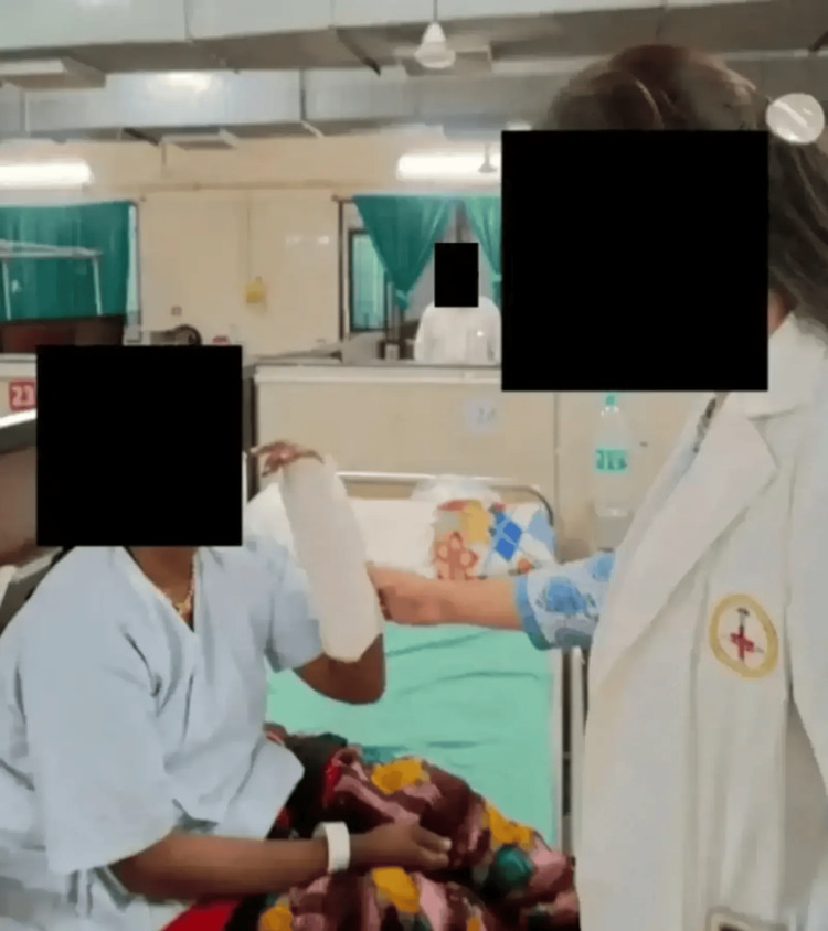 Figure 4
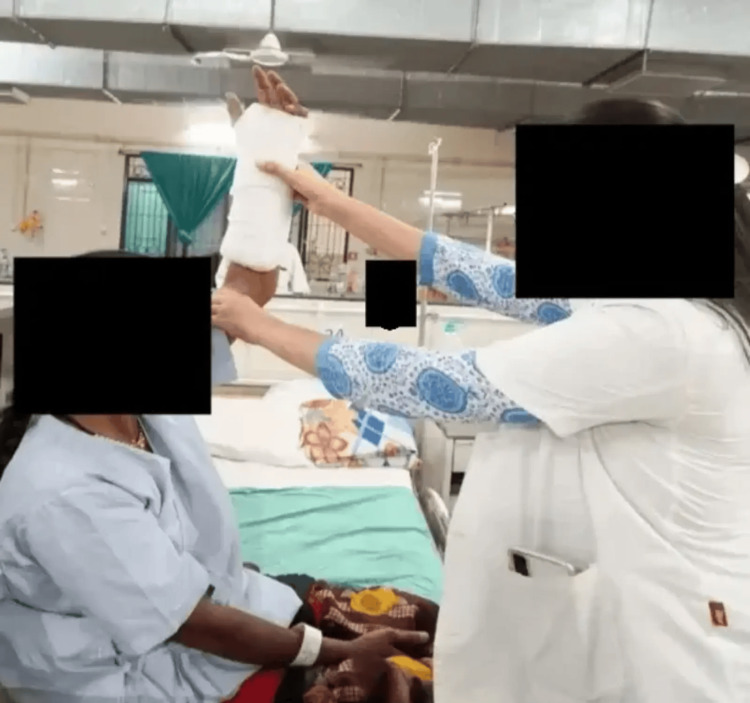 Figure 5
| Episode | Date of episode |
| Date of admission | 15/08/2023 |
| Date of operation | 17/08/2023 |
| Date of examination | 22/08/2023 |
| Goal | Intervention | Rationale |
| To educate the patient | Lifestyle modifications: to lift less weight with the right wrist, not to lift any heavy weight, and not to do any vigorous activities | To maintain health and increase well-being in life |
| To minimize pain and swelling | Use cryotherapy and elevate the affected limb | To promote a sense of well-being |
| To enhance wrist strength | Thera-band wrist exercises | To improve wrist range |
| To enhance wrist range of motion | Wrist flexion, wrist extension, ulnar deviation, and radial deviation | To improve wrist motion ranges |
| To improve functional activities of fingers | Opposition exercises | To improve finger mobility |
| To enhance shoulder range of motion | Shoulder flexion and extension, shoulder shrugs, and shoulder elevation and depression | To improve shoulder mobility and strength |
| To improve elbow range of motion | Elbow flexion and elbow extension | To improve elbow mobility and strength |
Peer Reviews
No public reviews on file for this paper yet. If you reviewed it on a platform where reviews are public (OpenReview, ICLR, NeurIPS, ICML), you can paste yours below so the community can read it here.
Videos
No videos yet. Explain this paper in a talk, walkthrough, or lecture? Add one.
Taxonomy
TopicsTraumatic Ocular and Foreign Body Injuries · Facial Trauma and Fracture Management · Child Abuse and Related Trauma
Introduction
The proximal hyperemic section of the joint is removed during a modified arthroplasty procedure for limited degenerative arthritis of the distal radioulnar joint, leaving the unaffected distal region intact [1]. Depending on the cubito-dorsal artery, a branch of the ulnar artery, the pisiform bone is vascularized. This procedure attempts to lessen discomfort, replace missing bones, maintain the space between the radius and capitatum and the scaphoid and triquetrum, and avoid carpal collapse. The vascularized relocation, which is suggested at stage III of conventional classifications, is verified using X-rays and MRI. The method entails employing ligament reconstructions to stabilize the transferred bone and may also include radiation shortening or scaphocapitate-restricted arthrodesis to lessen stress [2]. The "compromised" wrist's secondary consequences of the falling lunate include the following: at the radiolunate and mid-carpal articulations, the central column is deteriorating; the middle column's collapse; radial column collapse; proximal row instability; and, lastly, radial column degeneration [3]. Lunate fractures result in irregularities on the articular surfaces. The outcome is good if the fracture is repaired in a stable form. However, there is a chance that the central column will degenerate if there is secondary degeneration of the lunate facet and capitate with each other [4,5]. With a scaphocapitate fusion, the central column can be avoided because it cannot be rebuilt. On rare occasions, the capitate articular surface is unharmed, allowing for hemiarthroplasty [6,7]. Surgery known as limited wrist fusions is performed on individuals with carpus-related arthritis to reduce pain and maintain wrist motion. With the main objective of fusing damaged joints while preserving unaffected movement, the decision about motion-preserving surgery depends on the origin of the arthritis, involved joints, and spared joints. It is typical for the radial column to collapse in conjunction with central column degeneration or collapse [8,9]. If avascular fragments are shown to be the cause of synovitis or substantial widening on a sagittal CT scan, the lunate is excised. The surgeon must use caution to prevent ulnar translocation by not injuring the volar carpal ligaments, which are connected to the volar pieces [3]. The Sauvé-Kapandji treatment can treat ulnar impaction syndrome cases, enhance mobility in the distal radioulnar joint, and lessen pain. It works best for painful and limited forearm rotation following a distal radius fracture, together with an old dislocation or joint damage [10]. In order to restore function following the excision of the distal ulna, the radioulnar joint fusion technique is a rather uncommon treatment for patients with distal radioulnar joint instability. The treatment is frequently done after other attempts to maintain forearm rotation have failed, and the fusions frequently necessitate additional surgery to establish a union. To hasten healing, iliac bone grafts are frequently incorporated into the fusion region today [11].
Case presentation
Patient information
The patient was a 67-year-old female who was referred for physiotherapy with a primary complaint of pain in her right wrist. The patient states that she fell off a tractor and broke her right wrist. Pain is sudden in onset and severe in nature; it is non-radiating, shows no diurnal variation, and is non-progressive in nature. The patient gives an alleged history of slip and fall four years back sustaining an injury to her right wrist. At a private hospital in Amravati, the patient had gone through a proximal row carpectomy on the right side. Table 1 shows the timeline.
Clinical findings
The patient had a good sense of place, time, and people. On local examination, there was a healthy scar over the volar aspect of the right wrist, which was approximately 4×1 cm. Figure 1 and Figure 2 show the pre-operative and post-operative X-ray images, respectively.
Pre-operative X-rayX-ray showing the right wrist joint. X-ray was taken in antero-posterior view before the operation
Post-operative X-rayX-ray showing the right wrist joint. X-ray was taken in antero-posterior view and lateral view on 20 August 2023. It shows degenerative changes at the trapezoid and triquetral bones and degeneration and thinning of the triangular fibrocartilage with edema around the styloid process
Diagnostic intervention
Figure 3, Figure 4, and Figure 5 show the rehabilitation of the patient.
Finger oppositionPatient performing opposition movement of the fingers
Elbow flexionPatient performing flexion movement of the elbow
Shoulder flexionPatient performing flexion movement of the shoulder
Physiotherapy rehabilitation
Some goals of the treatment are to educate the patient, reduce her pain, and improve her range of motion, quality of life, and functional ability to do the daily household work.
Table 2 shows the physiotherapeutic protocol.
Discussion
Lunate fractures are uncommon and typically coexist with other fractures, ligament disruptions, or dislocations. In order to protect the lunate from shear and compression loads, the therapy entails decompressing it. While some lessen loads, they could put too much pressure on the lunate's ulnar side. Some procedures are extra-articular, protecting the architecture of the mid-carpal joint and the vascularization of the capsule [12]. Following proximal row carpectomy, immediate immobilization is not necessary, and early recovery is crucial. Early rehabilitation could shorten the time needed to regain strength and range of motion, as well as probably the time needed to resume work [13]. A common and effective surgical procedure for post-traumatic and degenerative radiocarpal and intercarpal articular lesions is proximal row carpectomy [14]. Activities for the range of motion were originally started passively and progressed to active-aided activities and then free workouts. There is proof that specialized exercise routines are important for reducing deficits and improving upper-limb function. After several sessions, heating has enhanced range of motion gain that is both acute and sustained in healthy people. In order to encourage motor point and muscle activity in an individual with limited hand function, an electrical muscle stimulant can be employed. Extensor muscle strength and motion range may increase functional abilities [15]. It is debatable whether the carpometacarpal joint should be included in TWA, i.e., total wrist arthrodesis, because difficulties at this location are reported in the literature [16]. When compared to patients with two distal screws, those who had several metacarpal screws had higher rates of plate-related problems and removal, but their post-operative grip strength was unaffected [17]. Patients who did not have fused index and long-ray carpometacarpal joints complained of pain, requiring a second arthrodesis treatment. Patients who perform substantial manual labor should have wrist arthrodesis performed together with index and long finger carpometacarpal joint arthroplasty [18]. Utilizing intramedullary pin fixation, a wrist arthrodesis technique has been enhanced, leading to successful fusion. This technique permits wrist placement at various degrees of ulnar deviation and extension while maintaining the speed and simplicity of earlier techniques [19]. A well-known operation that can be carried out using a variety of approaches is wrist arthrodesis. The most widely acknowledged posture for optimum hand function is between 10° and 30° of extension and 0° and 15° of ulnar deviation [20].
Conclusions
Proximal row carpectomy and partial carpal fusions serve as standard treatments for wrist arthritis, aiming to preserve some wrist motion while alleviating pain. However, outcomes can be unpredictable. In cases of pancarpal arthritis, total wrist fusion is another established option. The progression from arthrodesis to arthroplasty in other joints, such as the hip, knee, ankle, and shoulder, has influenced the evolving approach to managing wrist arthritis, with advancements contributing to more reliable treatment options. Physiotherapy helps reduce the pain and gain a functional range of motion allowing a good quality of life in such patients.
The reference list from the paper itself. Each links out to its DOI / PubMed record.
- 1Modified arthroplasty of the distal radio-ulnar joint J Hand Surg Br Watson HK Manzo RL 3223252720021216296810.1054/jhsb.2002.0805 · doi ↗ · pubmed ↗
- 2Vascularized pisiform transfer in place of lunatum for Kienböck's disease [Article in French]Chir Main Saffar P 0829201010.1016/j.main.2010.09.00521075664 · doi ↗ · pubmed ↗
- 3Kienböck disease: a new algorithm for the 21st century J Wrist Surg Lichtman DM Pientka WF 2nd Bain GI 210620172811979010.1055/s-0036-1593734 PMC 5258126 · doi ↗ · pubmed ↗
- 4Arthroscopic assessment and classification of Kienbock's disease Tech Hand Up Extrem Surg Bain GI Begg M 8131020061662811410.1097/00130911-200603000-00003 · doi ↗ · pubmed ↗
- 5An articular-based approach to Kienbock avascular necrosis of the lunate Tech Hand Up Extrem Surg Bain GI Durrant A 41471520112135852510.1097/BTH.0b 013e 31820 e 82e 8 · doi ↗ · pubmed ↗
- 6Distal radius hemiarthroplasty combined with proximal row carpectomy: case report Iowa Orthop J Boyer JS Adams B 168173302010 https://pubmed.ncbi.nlm.nih.gov/21045991/21045991 PMC 2958290 · pubmed ↗
- 7Decision making for partial carpal fusions J Wrist Surg Bain GI Mc Guire DT 103114120122417971310.1055/s-0032-1329548 PMC 3658677 · doi ↗ · pubmed ↗
- 8Avascular necrosis of the carpal lunate Clin Orthop Marek FM 96107101957 https://pubmed.ncbi.nlm.nih.gov/13561556/13561556 · pubmed ↗