Diagnostic and prognostic value of dual-point amyloid PET in Alzheimer’s disease (AD) mimickers
Luca Sofia, Federico Massa, Stefano Raffa, Matteo Pardini, Dario Arnaldi, Matteo Bauckneht, Silvia Morbelli

Abstract
Genes, proteins, chemicals, diseases, species, mutations and cell lines named across the full text — each resolved to its canonical identifier and authoritative record.
Click any figure to enlarge with its caption.
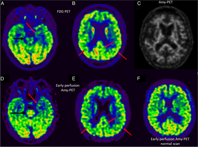 Figure 1
Peer Reviews
No public reviews on file for this paper yet. If you reviewed it on a platform where reviews are public (OpenReview, ICLR, NeurIPS, ICML), you can paste yours below so the community can read it here.
Videos
No videos yet. Explain this paper in a talk, walkthrough, or lecture? Add one.
Taxonomy
TopicsDementia and Cognitive Impairment Research · Alzheimer's disease research and treatments · Neurological Disease Mechanisms and Treatments
A 74-year-old man was referred to the neurologist for memory complaint. As prodromal AD was suspected, the patient underwent cerebrospinal fluid (CSF) biomarker evaluation which was not consistent with AD (negative for brain amyloidosis while mildly positive for both tau pathology and neurodegeneration: A-T + N +) [1]. To investigate non-AD underlying etiologies, the patient underwent a [^18^F]FDG-PET showing bilateral hypometabolism in the medial temporal lobes (MTL), posterior parietal cortex, and left posterior cingulate and precuneus (A, B). In the absence of brain amyloidosis, more prominent MTL hypometabolism suggested a preliminary categorization as suspected non-AD pathophysiology (SNAP) [2]. SNAP is a biomarker-based classification referring to individuals suggestive for AD-like neurodegeneration without β-amyloidosis. Tau pathology, as in primary age-related tauopathy (PART), has been hypothesized to play a major role in SNAP patients and was considered a possible etiology in this case [3]. However, the concomitant occurrence of hypometabolism in the posterior cortical regions, despite being previously described in SNAP patients due to disconnection from the hippocampus, could not allow exclusion of AD [2, 3]. Therefore, the patient underwent an amyloid-PET with [^18^F]Florbetaben which confirmed to be negative (A—according to the ATN system) (upper panel image C). Notably, amyloid-PET was acquired with a dual-point protocol including a short (5 min) image immediately after injection followed by a late steady-state standard acquisition [4]. Early acquisition is considered a perfusion-weighted phase representing a surrogate for neurodegeneration [4]. Early-perfusion amyloid PET imaging tightly mirrored the [^18^F]FDG-PET pattern (D, E; normal scan as a reference: F) [5]. Red arrows show regions of overlapping hypometabolism between [^18^F]FDG-PET and early-perfusion amyloid PET. Black arrow highlights the expected higher signal in brainstem on early-perfusion amyloid PET with respect to [^18^F]FDG. This case emphasizes the added value of dual-point amyloid-PET for the identification of AD mimickers with early perfusion imaging possibly replacing [^18^F]FDG-PET and providing prognostic stratification based on the extension/severity of neurodegeneration (although semiquantitive approaches to early-perfusion PET still need proper validation) [5]. Concomitant standard–late amyloid PET contributes to the exclusion of AD. Correlation between A/N status and topography of neurodegeneration might be relevant for diagnosis and prognosis also in AD mimickers.
The reference list from the paper itself. Each links out to its DOI / PubMed record.
- 1Jack CR Jr Bennett DA Blennow K Carrillo MC Dunn B Haeberlein SBNIA-AA research framework: toward a biological definition of Alzheimer’s disease Alzheimers Dement 201814453556210.1016/j.jalz.2018.02.01829653606 PMC 5958625 · doi ↗ · pubmed ↗
- 2Jack CR Jr Knopman DS Chételat G Dickson D Fagan AM Frisoni GB Suspected non-Alzheimer disease pathophysiology–concept and controversy Nat Rev Neurol 20161221172410.1038/nrneurol.2015.25126782335 PMC 4784257 · doi ↗ · pubmed ↗
- 3Jack CR Jr PART and SNAP Acta Neuropathol 2014128677377610.1007/s 00401-014-1362-325380757 PMC 4231211 · doi ↗ · pubmed ↗
- 4Florek L Tiepolt S Schroeter ML Berrouschot J Saur D Hesse S Dual time-point [18F]florbetaben PET delivers dual biomarker information in mild cognitive impairment and Alzheimer’s disease J Alzheimers Dis 20186631105111610.3233/jad-18052230400095 · doi ↗ · pubmed ↗
- 5Boccalini C Peretti DE Ribaldi F Scheffler M Stampacchia S Tomczyk S Early-phase 18F-Florbetapir and 18F-Flutemetamol images as proxies of brain metabolism in a memory clinic setting J Nucl Med 202264226627310.2967/jnumed.122.26425635863896 PMC 9902851 · doi ↗ · pubmed ↗
- 6Garibotto V Morbelli S Pagani M Dual-phase amyloid PET: hitting two birds with one stone Eur J Nucl Med Mol Imaging 20164391300130310.1007/s 00259-016-3393-627221519 · doi ↗ · pubmed ↗