Malignant gastric outlet obstruction: direct biopsy in the submucosal tunnel to obtain the diagnosis
Liansong Ye, Nuoya Zhou, Yi Mou, Chao Zhang, Lihong Wei, Xinhua Zhang, Bing Hu

Abstract
Genes, proteins, chemicals, diseases, species, mutations and cell lines named across the full text — each resolved to its canonical identifier and authoritative record.
Click any figure to enlarge with its caption.
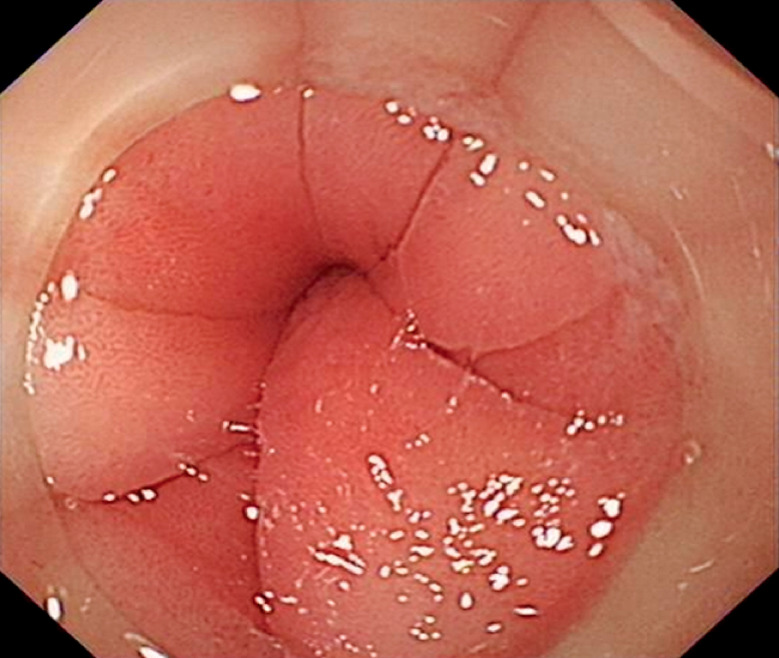 Fig. 1
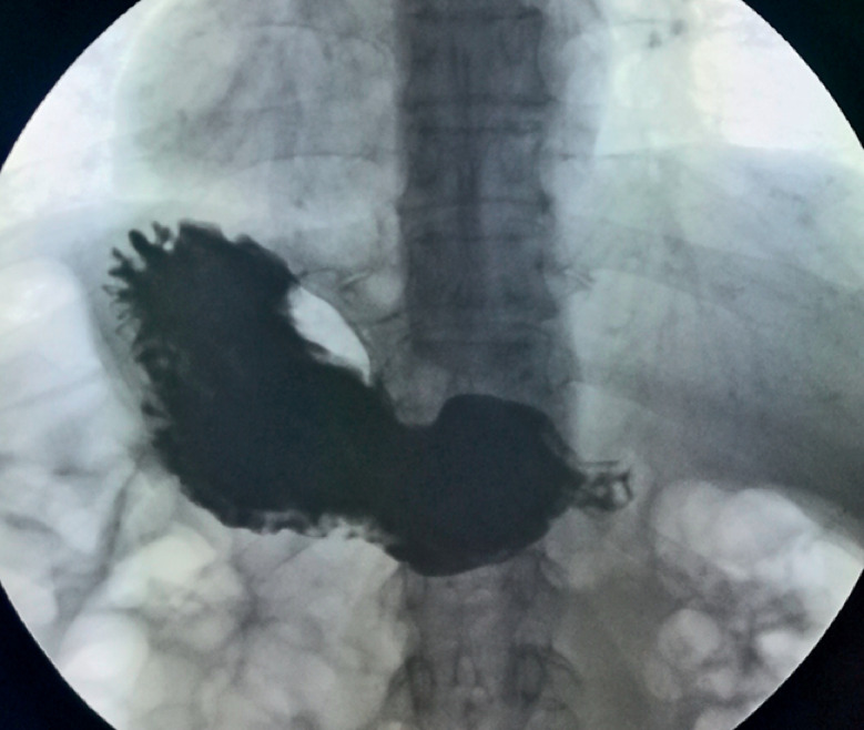 Fig. 2
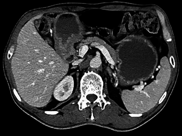 Fig. 3
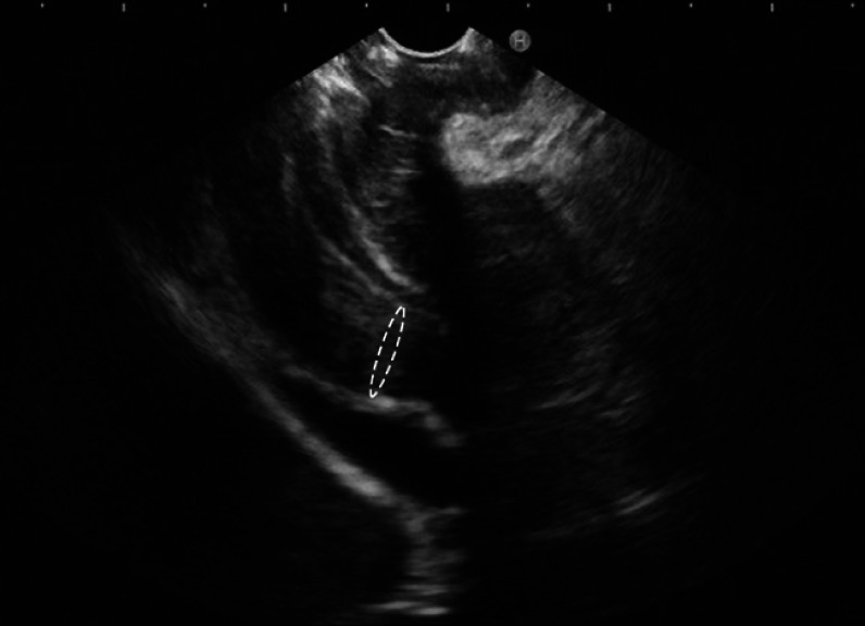 Fig. 4
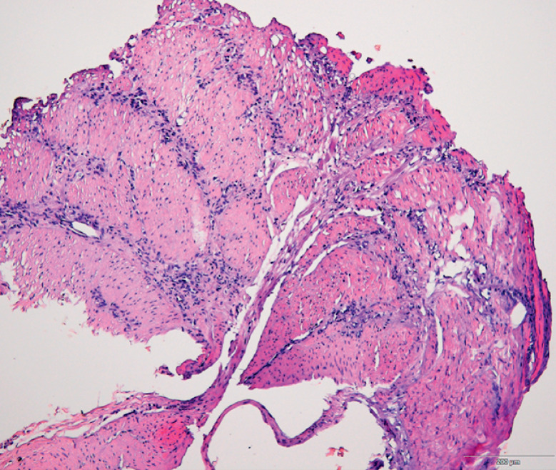 Fig. 5
- —Chengdu Science and Technology
- —China Postdoctoral Science Foundation10.13039/501100002858
- —Natural Science Foundation of Sichuan Province10.13039/501100018542
Peer Reviews
No public reviews on file for this paper yet. If you reviewed it on a platform where reviews are public (OpenReview, ICLR, NeurIPS, ICML), you can paste yours below so the community can read it here.
Videos
No videos yet. Explain this paper in a talk, walkthrough, or lecture? Add one.
Taxonomy
TopicsEsophageal and GI Pathology · Gastrointestinal disorders and treatments · Gastrointestinal Tumor Research and Treatment
A 59-year-old man presented with recurring early satiety for 1 year and postprandial vomiting for 2 months; he had also experienced weight loss of approximately 12 kg over the previous year. Gastroscopy showed food retention in the gastric cavity and an endoscope with a diameter of 8.9 mm could not be passed through the pylorus ( Fig. 1. Fig. 1 ). The mucosa of the pylorus appeared normal. A barium swallow showed delayed emptying of the stomach, with no filling defects or niches ( Fig. 2. Fig. 2 ). Abdominal computed tomography showed localized thickening of the gastric antrum, without enlargement of the lymph nodes ( Fig. 3. Fig. 3 ). Endoscopic ultrasonography (EUS) showed thickened muscularis propria at the pylorus ( Fig. 4. Fig. 4 ). Given the presumed diagnosis of hypertrophic pyloric stenosis, and after the patient had given informed consent, we performed peroral endoscopic myotomy (POEM) ( Video 1Video 1 ).
Fig. 1 Endoscopic image showing the normal-appearing mucosa of the pylorus, although the endoscope could not be passed through to the duodenum.
Fig. 2 Radiographic image from a barium swallow showing delayed emptying of the stomach, with no filling defects or niches.
Fig. 3 Abdominal computed tomography image showing localized thickening of the gastric antrum, with no enlarged lymph nodes.
Fig. 4 Endoscopic ultrasonography image showing thickening of the muscularis propria at the pylorus.
A direct biopsy is performed in the submucosal tunnel to obtain diagnostic tissue in a case of malignant gastric outlet obstruction.Video 1Video 1
Submucosal injection was performed on the posterior wall 8 cm proximal to the pylorus. A submucosal tunnel was subsequently created, but the procedure was interrupted because of dense adhesions of the thickened whitish muscularis propria and superficial mucosa. A sample of tissue from the thickened muscularis was obtained for pathology using a snare. Pathological findings subsequently showed the presence of atypical cells ( Fig. 5. Fig. 5 ), and immunohistochemistry demonstrated that these atypical cells were positive for PCK and CK8, confirming a poorly differentiated gastric adenocarcinoma. The patient underwent surgical intervention, followed by systemic chemotherapy, but tumor recurrence was detected within 1 year.
Fig. 5 Histological appearance of the biopsy of the thickened muscularis propria showing several atypical cells in the muscular tissue.
Gastric outlet obstruction caused by gastric carcinoma is common in clinical practice 11 ; however, in this case, the advanced gastric carcinoma did not present with one of the commonly seen growth patterns, such as a polypoid, fungating, ulcerating, or diffusely infiltrating lesion 22 , and the superficial mucosa above it was normal, which made the preoperative diagnosis difficult. Techniques such as the taking of deep samples via ESD that allow the full submucosa to be sampled could help with diagnosis. As per our experience, direct biopsy in the submucosal tunnel can also help make the final diagnosis.
Endoscopy_UCTN_Code_TTT_1AO_2AC
The reference list from the paper itself. Each links out to its DOI / PubMed record.
- 1Fukami N Anderson MA Khan K The role of endoscopy in gastroduodenal obstruction and gastroparesis Gastrointest Endosc 201174132110.1016/j.gie.2010.12.00321704805 · doi ↗ · pubmed ↗
- 2Hu B El Hajj N Sittler S Gastric cancer: Classification, histology and application of molecular pathology J Gastrointest Oncol 2012325126110.3978/j.issn.2078-6891.2012.02122943016 PMC 3418539 · doi ↗ · pubmed ↗