Endoscopic ultrasound-guided drainage for infected biloma using a unique long-type balloon catheter
Yuichi Takano, Naoki Tamai, Jun Noda, Tetsushi Azami, Fumitaka Niiya, Fumiya Nishimoto, Masatsugu Nagahama

Abstract
Genes, proteins, chemicals, diseases, species, mutations and cell lines named across the full text — each resolved to its canonical identifier and authoritative record.
Click any figure to enlarge with its caption.
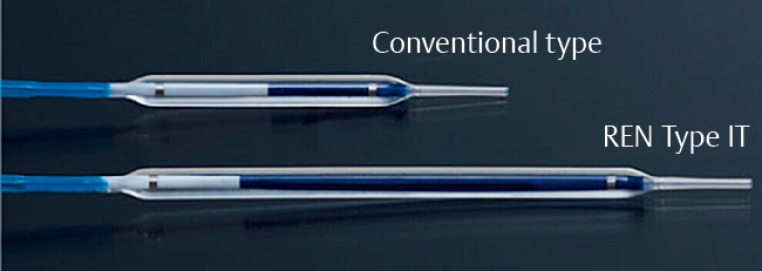 Fig. 1
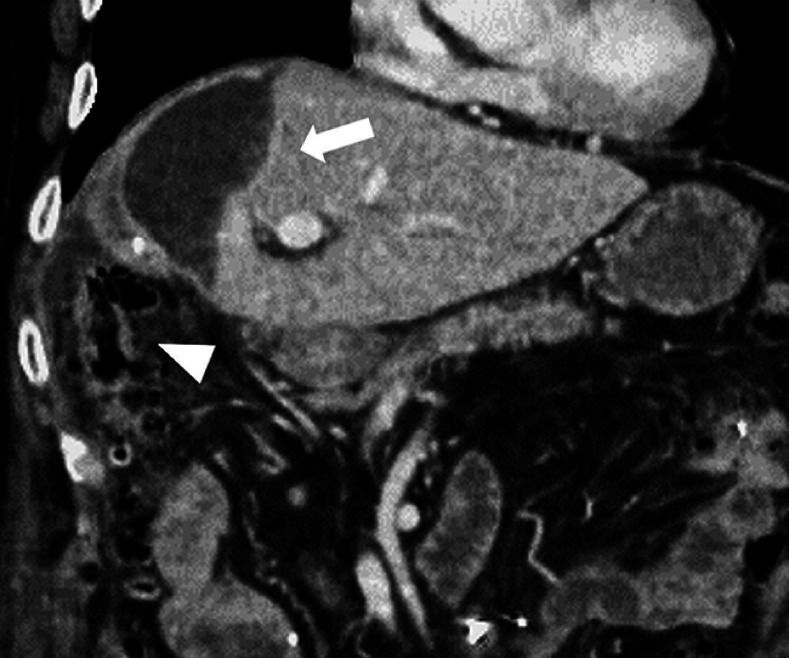 Fig. 2
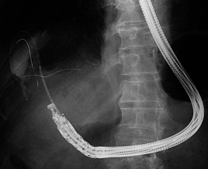 Fig. 3
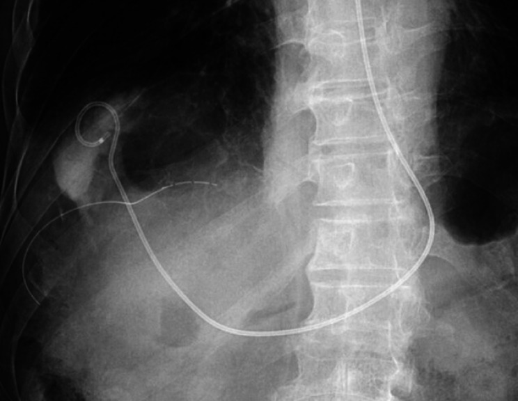 Fig. 4
Peer Reviews
No public reviews on file for this paper yet. If you reviewed it on a platform where reviews are public (OpenReview, ICLR, NeurIPS, ICML), you can paste yours below so the community can read it here.
Videos
No videos yet. Explain this paper in a talk, walkthrough, or lecture? Add one.
Taxonomy
TopicsGallbladder and Bile Duct Disorders · Esophageal and GI Pathology · Pediatric Hepatobiliary Diseases and Treatments
Endoscopic ultrasound (EUS)-guided drainage is a widely performed procedure. However, tract dilation can sometimes present challenges and many dedicated devices have been developed 11 22 33 . Herein, we describe a case of successful tract dilation using a unique long-type balloon catheter ( Fig. 1. Fig. 1 ).
Fig. 1 A unique long-type balloon catheter of 3 mm in diameter and 6 cm in length (REN Type IT; Kaneka). This balloon is twice as long as the 3 cm-length conventional type (REN, Kaneka).
A 77-year-old man who underwent open extended right hepatectomy, extrahepatic bile duct resection, and choledochojejunostomy for gallbladder cancer developed abdominal pain and fever 15 days postoperatively. Contrast-enhanced computed tomography revealed fluid collection under the right diaphragm, suggesting an infected biloma. Due to the position of the ascending colon in the puncture line, percutaneous drainage was not possible ( Fig. 2. Fig. 2 ). As a result, EUS-guided drainage was selected.
Fig. 2 Contrast-enhanced computed tomography demonstrates fluid collection under the right diaphragm, suggesting an infected biloma (arrow). The ascending colon is present in the puncture line (arrowhead), making percutaneous drainage impossible.
An echoendoscope (GF-UCT260; Olympus Medical Systems, Tokyo, Japan) was inserted into the duodenal bulb and twisted counterclockwise, revealing a biloma measuring 58 × 43 mm. The distance from the echoendoscope to the biloma was 46 mm. A 19G needle (EZshot3, Olympus Medical Systems) was used to puncture through the liver parenchyma. After injecting contrast medium, a 0.025-inch guidewire (Visiglide2, Olympus Medical Systems) was placed. A balloon catheter with a diameter of 3 mm and length of 6 cm (REN Type IT; Kaneka, Tokyo, Japan) was inserted smoothly, and tract dilation was performed. The expanded balloon was clearly visible on the fluoroscopic image ( Fig. 3. Fig. 3 ). Finally, a 6-Fr pigtail-type endoscopic nasobiliary drainage (NB-Braid; Piolax, Yokohama, Japan) was placed, and the procedure was completed without any adverse events ( Fig. 4. Fig. 4 , Video 1Video 1 ).
Fig. 3 Tract dilation was performed with a balloon catheter of 3 mm in diameter and 6 cm in length (REN Type IT, Kaneka). The expanded balloon was easily recognized on the fluoroscopic image.
Fig. 4 A 6-Fr pigtail-type endoscopic nasobiliary drainage (NB-Braid, Piolax) was placed.
Endoscopic ultrasound-guided drainage for infected biloma using a novel long-type balloon catheter.Video 1Video 1
In this case, a conventional-length balloon catheter would require multiple dilations. The use of this long-type balloon catheter allowed simultaneous dilation of the duodenal mucosa and liver parenchyma in a single inflation. This device can simplify tract dilation and shorten procedure time.
Endoscopy_UCTN_Code_TTT_1AS_2AD
The reference list from the paper itself. Each links out to its DOI / PubMed record.
- 1Honjo M Itoi T Tsuchiya T Safety and efficacy of ultra-tapered mechanical dilator for EUS-guided hepaticogastrostomy and pancreatic duct drainage compared with electrocautery dilator (with video)Endosc Ultrasound 2018737638229882518 10.4103/eus.eus_2_18PMC 6289009 · doi ↗ · pubmed ↗
- 2Okuno N Hara K Haba S Novel drill dilator facilitates endoscopic ultrasound-guided hepaticogastrostomy Dig Endosc 20233538939310.1111/den.1444736170547 · doi ↗ · pubmed ↗
- 3Iwashita T Ogura T Ishiwatari H Utility of dedicated bougie dilator for a 0.018-inch guidewire during EUS-guided biliary drainage: A multi-center retrospective cohort study J Hepatobiliary Pancreat Sci 20222981081634272831 10.1002/jhbp.1021 · doi ↗ · pubmed ↗