Correction: Blastulation time measured with time-lapse system can predict in vitro viability of bovine blastocysts
Carmen Huayhua, Misael Rodríguez, Jhorjhi Vega, Mario Briones, Lleretny Rodriguez-Alvarez, Edwin Mellisho

Abstract
Genes, proteins, chemicals, diseases, species, mutations and cell lines named across the full text — each resolved to its canonical identifier and authoritative record.
Click any figure to enlarge with its caption.
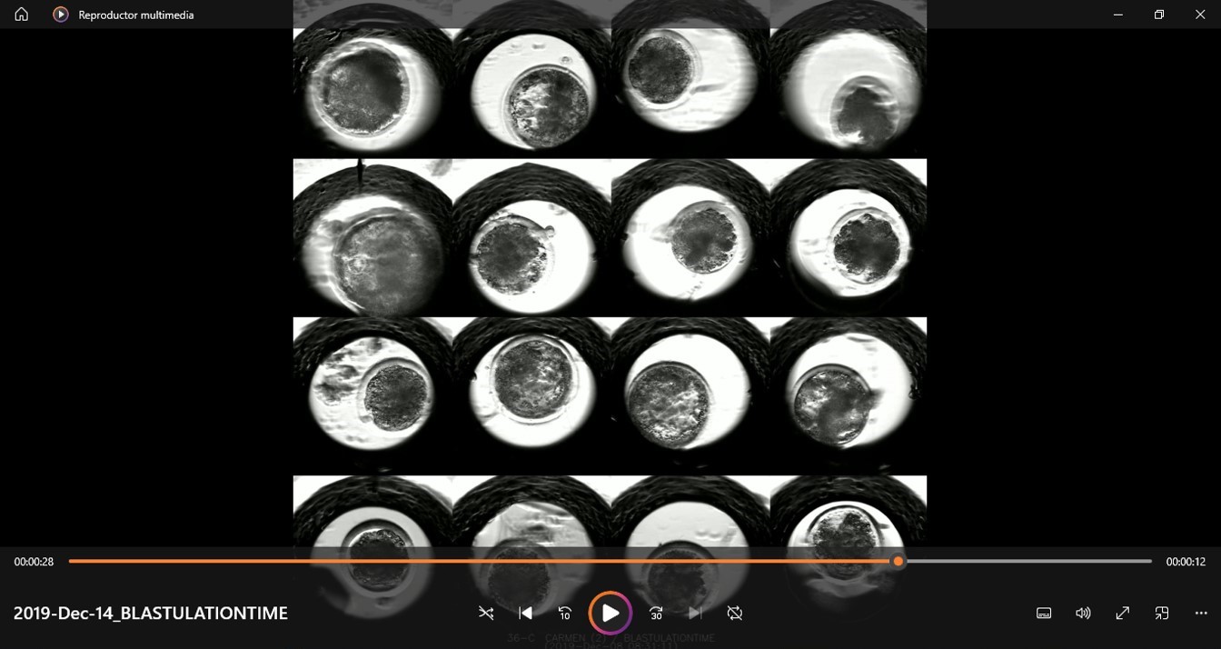 Figure 1
 Figure 2
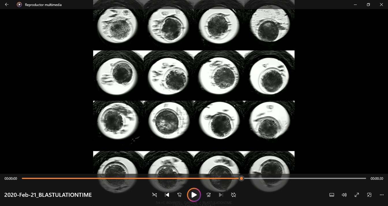 Figure 3
Peer Reviews
No public reviews on file for this paper yet. If you reviewed it on a platform where reviews are public (OpenReview, ICLR, NeurIPS, ICML), you can paste yours below so the community can read it here.
Videos
No videos yet. Explain this paper in a talk, walkthrough, or lecture? Add one.
Taxonomy
TopicsReproductive Biology and Fertility · Pluripotent Stem Cells Research · Renal and related cancers
After publication of this article [1], concerns were raised that Fig 2 in [1] was duplicated from Figure 3 in a previous article [2] by a different author group, and that [2] was not cited in [1].
During editorial follow up on this issue, the corresponding author stated that Fig 2 in [1] was included in error.
Owing to the concerns about duplication with previously published content [2], published in 2010 by Oxford University Press [2], which is not offered under a CC BY license, this article [1] was republished on June 24, 2024, to correct the Fig 2 error. The updated figure reports demonstrative images of individual cultures in 16 microwell plates to monitor embryonic development in cattle. These images are from the original experiments reported in [1] at day 7.5 post IVF (S5 File). Replicate images from the time of the original experiments and images from earlier and later repeat experiments are provided here in S1–S4 Files. Additional data underlying S1 Table and supporting the embryo measurements in [1] are provided here in S6–S15 Files.
An member of the Editorial Board reviewed the concerns and stated that the updated Fig 2 supports what is stated in the published article, ‘During in vitro culture, 16 presumptive zygotes were individually (Fig 2) monitored using the Primo Vision TL equipment (Vitrolife, Sweden) that takes images every 15 minutes from day 1 to 7.5 post IVF.’ [1]. They also advised that the data provided with this notice are sufficient to support the article’s key results as required by the PLOS Data Availability policy.
The corresponding author also provided some clarifying information about the methodology used in [1]:
Supporting information
S1 FileImages from earlier repeat experiments supporting Fig 2.(JPG)
S2 FileImages from later repeat experiments supporting Fig 2.(JPG)
S3 FileImages from earlier repeat experiments supporting Fig 2.(JPG)
S4 FileReplicate images supporting Fig 2 from the time of the original experiments.(PNG)
S5 FileImages supporting Fig 2 from the time of the original experiments.(PNG)
S6 FileUnderlying data supporting the summary in S1 Table in [1].(XLSX)
S7 FileSupporting data for S1 Table and the embryo measurements in [1].The times to divisions 2, 4, 8, etc. were processed individually, and the kinematics of the embryo was recorded in two parts because the Primovision (R) software did not allow continuous video recording for the 7.5 days post IVF. S7–S13 Files correspond to three batches of work.(WEBM)
S8 FileSupporting data for S1 Table and the embryo measurements in [1].The times to divisions 2, 4, 8, etc. were processed individually, and the kinematics of the embryo was recorded in two parts because the Primovision (R) software did not allow continuous video recording for the 7.5 days post IVF. S7–S13 Files correspond to three batches of work.(WEBM)
S9 FileSupporting data for S1 Table and the embryo measurements in [1].The times to divisions 2, 4, 8, etc. were processed individually, and the kinematics of the embryo was recorded in two parts because the Primovision (R) software did not allow continuous video recording for the 7.5 days post IVF. S7–S13 Files correspond to three batches of work.(WEBM)
S10 FileSupporting data for S1 Table and the embryo measurements in [1].The times to divisions 2, 4, 8, etc. were processed individually, and the kinematics of the embryo was recorded in two parts because the Primovision (R) software did not allow continuous video recording for the 7.5 days post IVF. S7–S13 Files correspond to three batches of work.(WEBM)
S11 FileSupporting data for S1 Table and the embryo measurements in [1].The times to divisions 2, 4, 8, etc. were processed individually, and the kinematics of the embryo was recorded in two parts because the Primovision (R) software did not allow continuous video recording for the 7.5 days post IVF. S7–S13 Files correspond to three batches of work.(WEBM)
S12 FileSupporting data for S1 Table and the embryo measurements in [1].The times to divisions 2, 4, 8, etc. were processed individually, and the kinematics of the embryo was recorded in two parts because the Primovision (R) software did not allow continuous video recording for the 7.5 days post IVF. S7–S13 Files correspond to three batches of work.(WEBM)
S13 FileSupporting data for S1 Table and the embryo measurements in [1].The times to divisions 2, 4, 8, etc. were processed individually, and the kinematics of the embryo was recorded in two parts because the Primovision (R) software did not allow continuous video recording for the 7.5 days post IVF. S7–S13 Files correspond to three batches of work.(WEBM)
S14 FileAn example of how morphokinetics parameters were assessed in [1].(PNG)
S15 FileAn embryo image of 9.5 post IVF (individual embryo culture) used to assess post-hatching development (in vitro viability).(PNG)
The reference list from the paper itself. Each links out to its DOI / PubMed record.
- 1Huayhua C, Rodríguez M, Vega J, Briones M, Rodriguez-Alvarez L, Mellisho E (2023) Blastulation time measured with time-lapse system can predict in vitro viability of bovine blastocysts. P Lo S ONE 18(8): e 0289751. doi: 10.1371/journal.pone.0289751 37561791 PMC 10414680 · doi ↗ · pubmed ↗
- 2Sugimura Satoshi, Akai Tomonori, Somfai Tamás, Hirayama Muneyuki, Aikawa Yoshio, Ohtake Masaki, Hattori Hideshi, Kobayashi Shuji, Hashiyada Yutaka, Konishi Kazuyuki, Imai Kei, Time-Lapse Cinematography-Compatible Polystyrene-Based Microwell Culture System: A Novel Tool for Tracking the Development of Individual Bovine Embryos, Biology of Reproduction, Volume 83, Issue 6, 1 December 2010, Pages 970–978, doi: 10.1095/biolreprod.110.085522 20739661 · doi ↗ · pubmed ↗