Dermoscopy of nasal and auricular gouty tophi
Bruno Simão dos Santos, Maria Augusta Pires Maciel, Neusa Yuriko Sakai Valente

Abstract
Genes, proteins, chemicals, diseases, species, mutations and cell lines named across the full text — each resolved to its canonical identifier and authoritative record.
Click any figure to enlarge with its caption.
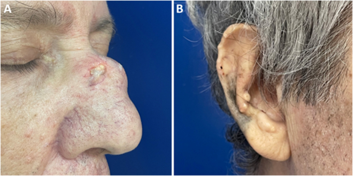 Figure 1
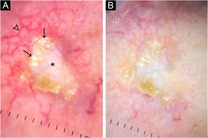 Figure 2
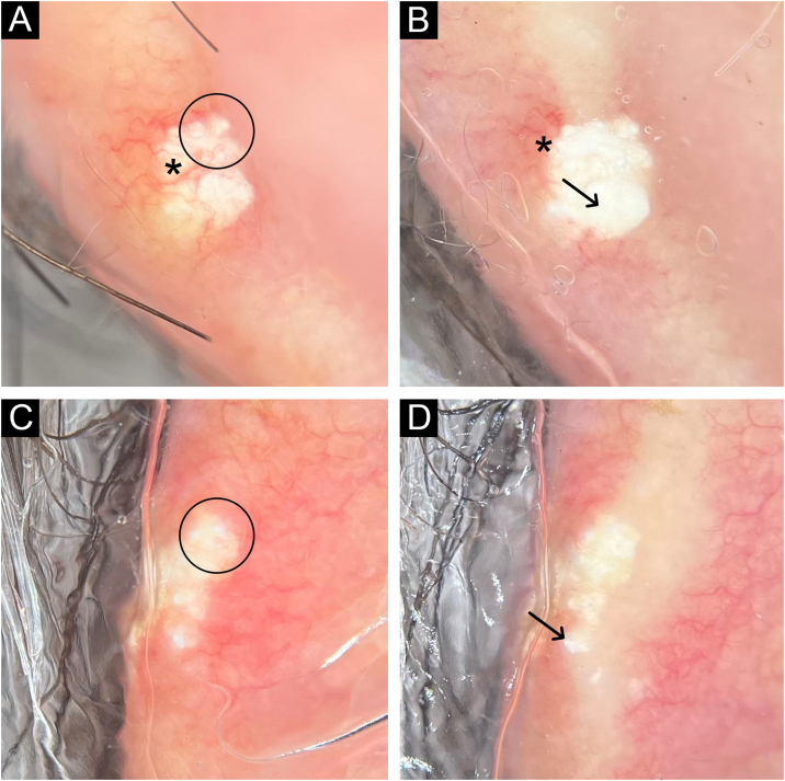 Figure 3
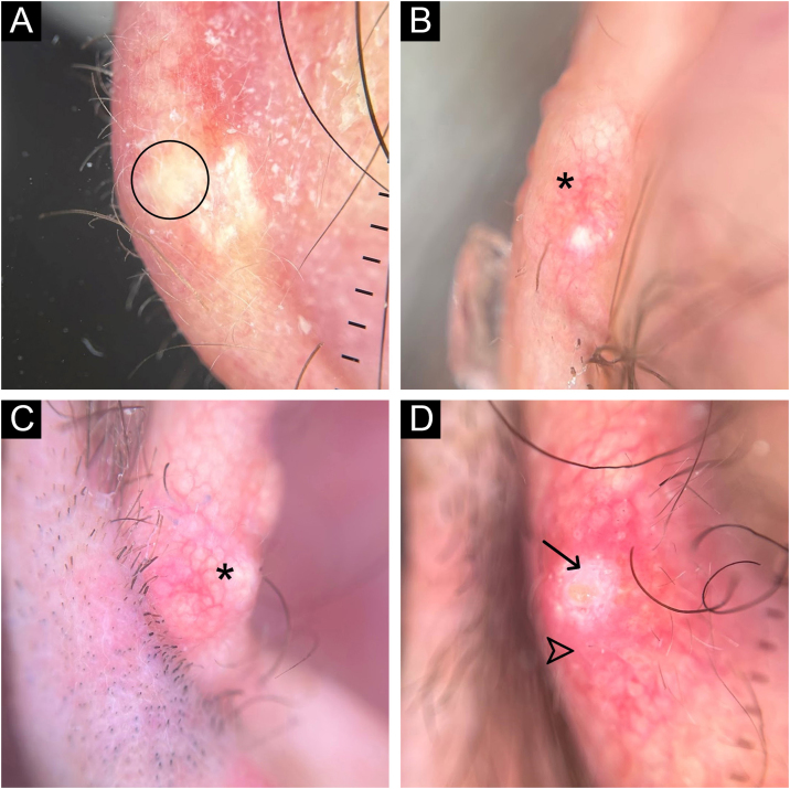 Figure 4
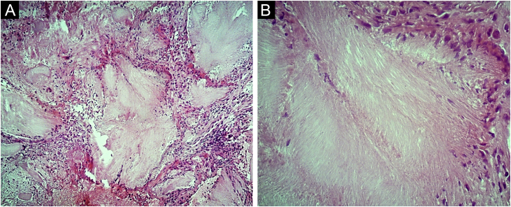 Figure 5
Peer Reviews
No public reviews on file for this paper yet. If you reviewed it on a platform where reviews are public (OpenReview, ICLR, NeurIPS, ICML), you can paste yours below so the community can read it here.
Videos
No videos yet. Explain this paper in a talk, walkthrough, or lecture? Add one.
Taxonomy
TopicsGout, Hyperuricemia, Uric Acid · Urticaria and Related Conditions · Bartonella species infections research
Dear Editor,
A 62-year-old male hypertensive patient, a former alcoholic, suffering from gout for approximately 20 years and undergoing irregular treatment with allopurinol and colchicine, presented with a firm and painless nodular lesion on the nasal dorsum for one year, which progressed with ulceration. On dermatological examination, yellowish papules on the ear helices (Fig. 1) and increased volume in the joints of the hands, elbows, knees and feet were also observed. Dermoscopy of the nasal lesion showed a central amorphous white area, and yellowish areas interspersed with shiny white polymorphic structures on the periphery of the lesion, in addition to diffuse erythema and peripheral branched vessels (Fig. 2). Dermoscopic examination of the helix lesions showed, predominantly, aggregated yellowish-white globular structures (Fig. 3), with branched vessels crossing the lesion and on its periphery (Fig. 3A). In other lesions of the right helix, unlike the previous findings, an amorphous yellowish-white area was observed (Fig. 4A) or an amorphous yellowish-white background with blurred branched vessels scattered over the lesion (Fig. 4B-C). Also in the same location, a lesion with an amorphous white area, a yellowish center and peripheral diffuse erythema could be observed, similar to the nasal lesion (Fig. 4D). The laboratory tests showed the patient had anemia and elevated inflammatory markers, reduced renal function and elevated serum uric acid levels (7.8 mg/dL, RV: 3.5‒7.2 mg/dL). However, urinary uric acid was within normal range (378.4 mg/24h ‒ RV: 250 to 750 mg/24h). Histopathological examination of the lesion on the nasal dorsum showed amorphous or crystalloid eosinophilic deposits in the dermis with a needle-like appearance, corresponding to aggregates of monosodium urate crystals, surrounded by a granulomatous inflammatory infiltrate, compatible with the diagnosis of gouty tophus (Fig. 5).Figure 1. Clinical aspect of gouty tophi. Ulcerated nodular lesion on the nasal dorsum (A) and yellowish papules on the right ear (B).Figure 1. Figure 2Dermoscopy with polarized light (A) and (B) of the nasal lesion. Central amorphous white area (asterisk), with yellowish areas interspersed with shiny white polymorphic structures (arrows) on the periphery of the lesion, diffuse erythema and blurred branched vessels (arrowhead) best seen in (A) due to the lack of contact between the dermatoscope and the skin. Original magnification, ×10.Figure 2. Figure 3Dermoscopy with polarized light (A to D) of two lesions on the helix. Aggregated yellowish-white globular structures (circles), with branched vessels (asterisks) crossing the lesion (A) and on its periphery (A and B). In the lesion shown in (C) and (D) there are no vessels over the lesion. Shiny white structures can be seen in both lesions (arrows). Original magnification, ×10.Figure 3. Figure 4Polarized light dermoscopy without contact with the helix lesions. In (A), amorphous yellowish-white area without vessels (circle); (B) and (C), yellowish-white background with blurred branched vessels over the lesion (asterisks); (D), amorphous white area (arrow) with a yellowish center and peripheral diffuse erythema (arrowhead). Original magnification, ×10.Figure 4. Figure 5Photomicrographs of the histopathology of the nasal dorsum lesion. Eosinophilic, amorphous or crystalloid deposits in the dermis, surrounded by a granulomatous inflammatory infiltrate. Hematoxylin & eosin, ×100 (A) and ×400 (B).Figure 5
Discussion
Gout is the most common inflammatory arthritis and is caused by the deposit of monosodium urate crystals in the joints.1 Gouty tophus, the accumulation of these crystals in soft tissues, is the characteristic clinical manifestation of advanced disease but may be the first clinical sign in some cases.2 On the skin, it is characterized by firm papules and nodules, with a smooth or multilobulated outline, normochromic, yellowish, or erythematous, which may be ulcerated. The most common locations include the first and fifth metatarsophalangeal joints and the hand and wrist joints. The presentation of gout in the head and neck region is uncommon. The nasal region is usually a rarely affected area.3, 4, 5
Regarding the dermoscopy of gouty tophus, Yoshida et al. reported the dermoscopic findings of an ulcerated gouty tophus on the right toe, describing the presence of whitish structures similar to “horns”, with some shiny dots.6 In the present case, grouped amorphous and globular white and yellowish-white areas were observed, associated with several shiny white structures of different shapes, observed both on polarized and non-polarized light. It is possible that such shiny structures correspond to accumulations of monosodium urate crystals located more superficially in the skin. Moreover, the lesions showed different dermoscopic findings compared to those reported in the previous published study.
The diagnosis of gouty tophus, in general, is based on clinicopathological correlation and there are few reports describing the dermoscopic findings of this clinical manifestation. Over the last few years, several studies have shown that dermoscopy can be useful in assisting the non-invasive diagnosis of various inflammatory and infectious diseases.7, 8, 9 Therefore, knowledge of the dermoscopic structures present in gouty tophi becomes relevant, as it can help in the differential diagnosis of dermatoses with a similar clinical picture, such as malignant neoplasms and other metabolic and storage diseases.
Financial support
None declared.
Authors’ contributions
Bruno Simão dos Santos: Design and planning of the study; drafting and editing of the manuscript; critical review of intellectual content; effective participation in research orientation; intellectual participation in the propaedeutic and/or therapeutic conduct of the studied case; critical review of the literature; approval of the final version of the manuscript.
Maria Augusta Pires Maciel: Drafting and editing of the manuscript; data collection; intellectual participation in the propaedeutic and/or therapeutic conduct of the studied case; critical review of the literature; approval of the final version of the manuscript.
Neusa Yuriko Sakai Valente: Critical review of intellectual content; effective participation in research orientation; intellectual participation in the propaedeutic and/or therapeutic conduct of the studied case; approval of the final version of the manuscript.
Conflicts of interest
None declared.
The reference list from the paper itself. Each links out to its DOI / PubMed record.
- 1Klauser A.S.Halpern E.J.Strobl S.Gruber J.Feuchtner G.Bellmann-Weiler R.Dual-energy computed tomography detection of cardiovascular monosodium urate deposits in patients with gout JAMA Cardiol 42019101910283150915610.1001/jamacardio.2019.3201 PMC 6739730 · doi ↗ · pubmed ↗
- 2Tognetti L.Cinotti E.Fiorani D.Rubegni P.Perrot J.L.Noninvasive diagnosis of liquefied gouty tophus: reflectance confocal microscopy as an alternative to polarizing light microscopy analysis Skin Res Technol 2520192402413032505710.1111/srt.12627 · doi ↗ · pubmed ↗
- 3Chen S.L.Chen J.R.Yang S.W.Painless gouty tophus in the nasal bridge: a case report and literature review Medicine (Baltimore)982019 e 148503088268210.1097/MD.0000000000014850 PMC 6426609 · doi ↗ · pubmed ↗
- 4Rottmann E.Bulbin D.Zaklama A.Gouty tophus erodes nasal bone Clin Rheumatol 4120229399413459971510.1007/s 10067-021-05897-z PMC 8873052 · doi ↗ · pubmed ↗
- 5Shiu W.H.L.Cheng H.M.J.Chan Y.T.Chu C.Y.Kan W.K.Gouty tophus: unusual case of nasal lump Radiol Case Rep 162021290429073440102210.1016/j.radcr.2021.06.094PMC 8350013 · doi ↗ · pubmed ↗
- 6Yoshida Y.Yamamoto O.Dermoscopic features of ulcerated gouty tophus Eur J Dermatol 19200964610.1684/ejd.2009.078919709981 · doi ↗ · pubmed ↗
- 7Errichetti E.Stinco G.Dermoscopy in general dermatology: a practical overview Dermatol Ther (Heidelb)620164715072761329710.1007/s 13555-016-0141-6PMC 5120630 · doi ↗ · pubmed ↗
- 8Bakos R.M.Reinehr C.Escobar G.F.Leite L.L.Dermoscopy of skin infestations and infections (entomodermoscopy) - Part I: dermatozoonoses and bacterial infections An Bras Dermatol 9620217357453462052410.1016/j.abd.2021.04.007PMC 8790189 · doi ↗ · pubmed ↗