Cinematic rendering of osteopoikilosis
Liwei Fu, Chong Tian, Xianchun Zeng

Abstract
Genes, proteins, chemicals, diseases, species, mutations and cell lines named across the full text — each resolved to its canonical identifier and authoritative record.
Click any figure to enlarge with its caption.
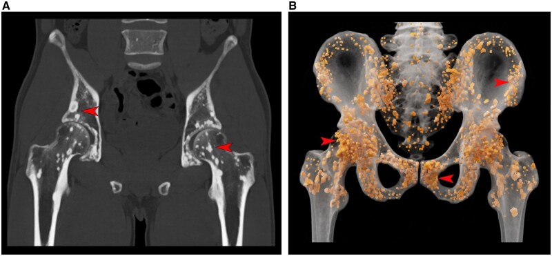 Figure 1
- —Guizhou Province Multi-level Innovative Talent Training Program
Peer Reviews
No public reviews on file for this paper yet. If you reviewed it on a platform where reviews are public (OpenReview, ICLR, NeurIPS, ICML), you can paste yours below so the community can read it here.
Videos
No videos yet. Explain this paper in a talk, walkthrough, or lecture? Add one.
Taxonomy
TopicsDermatological and Skeletal Disorders · Dupuytren's Contracture and Treatments · Medicine and Dermatology Studies History
A 26-year-old man presented to the urology department with ejaculatory dysfunction. The patient reported a history of diabetes, and apart from elevated levels of plasma glucose and glycated haemoglobin in the laboratory tests, no other abnormalities were observed. Pelvic CT revealed diffuse dense shadows in the pelvic region and bilateral proximal femurs (Fig. 1A), which were further highlighted using a cinematic rendering reconstruction technique (Fig. 1B and Supplementary Video, available at Rheumatology online. After supplementary genetic testing revealed the presence of a LEMD3 gene mutation in the patient, the physician made a clinical diagnosis of osteopoikilosis based on the combined assessment of the CT examination results.
Osteopoikilosis is a rare asymptomatic osteosclerotic dysplasia, with an incidence of 1 in 50 000 people [1]. Most scholars believe that this condition is associated with mutations in the LEMD3 gene. These lesions are most commonly discovered incidentally on radiographic imaging, and they are symmetric in nature but with an uneven distribution. Compared with conventional CT images, cinematic rendering can apply different transfer functions for specific cases and structures, aiding more intuitive visualization of spot extent in one view [2].
Supplementary Material
keae234_Supplementary_Data
The reference list from the paper itself. Each links out to its DOI / PubMed record.
- 1Perin S , Rabach I, Pascolo P et al A spotted bone. J Pediatr 2016;176:220–220.e 1.27301574 10.1016/j.jpeds.2016.05.069 · doi ↗ · pubmed ↗
- 2Eid M , De Cecco CN, Nance Jr JW, et al Cinematic rendering in CT: a novel, lifelike 3D visualization technique. AJR Am J Roentgenol 2017;209:370–9.28504564 10.2214/AJR.17.17850 · doi ↗ · pubmed ↗