Pleuropulmonary blastoma: a rare clinical image
Ashwin Karnan

Abstract
Genes, proteins, chemicals, diseases, species, mutations and cell lines named across the full text — each resolved to its canonical identifier and authoritative record.
Click any figure to enlarge with its caption.
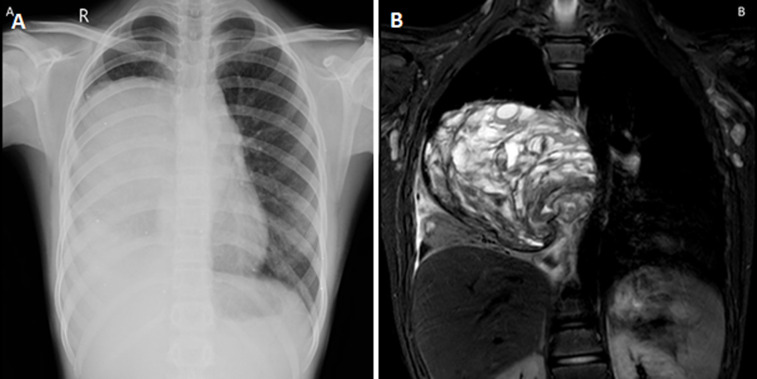 Figure 1
Peer Reviews
No public reviews on file for this paper yet. If you reviewed it on a platform where reviews are public (OpenReview, ICLR, NeurIPS, ICML), you can paste yours below so the community can read it here.
Videos
No videos yet. Explain this paper in a talk, walkthrough, or lecture? Add one.
Taxonomy
TopicsCongenital Diaphragmatic Hernia Studies · Tracheal and airway disorders · Esophageal and GI Pathology
Image in medicine
A 13-year-old boy presented to the outpatient department with complaints of breathing difficulty for the past 2 months. Magnetic resonance imaging of the thorax showed a solid cystic mass lesion in the right hemithorax of size 8.9 x 10.7 cm with mild pleural effusion with mass effect shifting the major vessels to the left side. Computed tomography-guided biopsy was done which showed variable thickened nodule-like areas with both single cells and cohesive aggregates with positive stains for vimentin and cytokeratin. A diagnosis of pleuropulmonary blastoma was made. The patient underwent surgical resection and is currently on follow-up. Pleuropulmonary blastomas are rare and aggressive childhood intrathoracic tumors common in children less than 6 years of age. It may be of three types- type I (cystic), type II (mixed), or type III (solid). Clinical presentation includes shortness of breath, chest pain, cough, and hemoptysis. Tumor size >5 cm with pleural or mediastinal invasion has a poor prognosis. Surgical resection, postoperative radiotherapy, and chemotherapy are available.
A) chest X-ray of the patient showing homogenous opacity in the right lung; B) MRI of the thorax showing solid cystic lesion in the right hemithorax with mild pleural effusion