Use of Three-Dimensional Echocardiography to Identify an Unusual Cause of Aortic Regurgitation
J. Kyle Buck, Manrique Alvarez, Sneha Chebrolu, Rohesh J. Fernando, Karl Richardson, Adrian L. Lata, Scott R. Coleman

TL;DR
This paper describes how 3D echocardiography helped identify a rare cause of aortic regurgitation following a heart procedure.
Contribution
The study highlights the diagnostic value of 3D imaging in identifying aortic valve injury when 2D imaging is inconclusive.
Findings
New-onset aortic regurgitation after coronary angiography may indicate aortic valve injury.
3D imaging revealed a partial tear in the right coronary cusp that 2D imaging missed.
TEE findings influenced changes in the patient's medical treatment.
Abstract
•New-onset AR after coronary angiography should raise suspicion for AV injury.•Consider 3D imaging if no clear etiology found with 2D imaging.•Three-dimensional imaging showed a partial tear of RCC.•Medical intervention was altered based in part on TEE findings. New-onset AR after coronary angiography should raise suspicion for AV injury. Consider 3D imaging if no clear etiology found with 2D imaging. Three-dimensional imaging showed a partial tear of RCC. Medical intervention was altered based in part on TEE findings.
Genes, proteins, chemicals, diseases, species, mutations and cell lines named across the full text — each resolved to its canonical identifier and authoritative record.
Click any figure to enlarge with its caption.
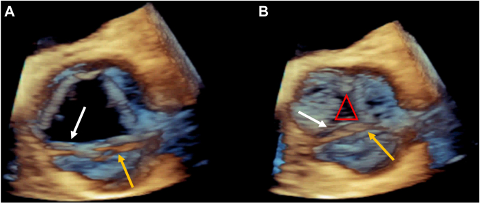 Figure 1
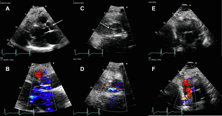 Figure 2
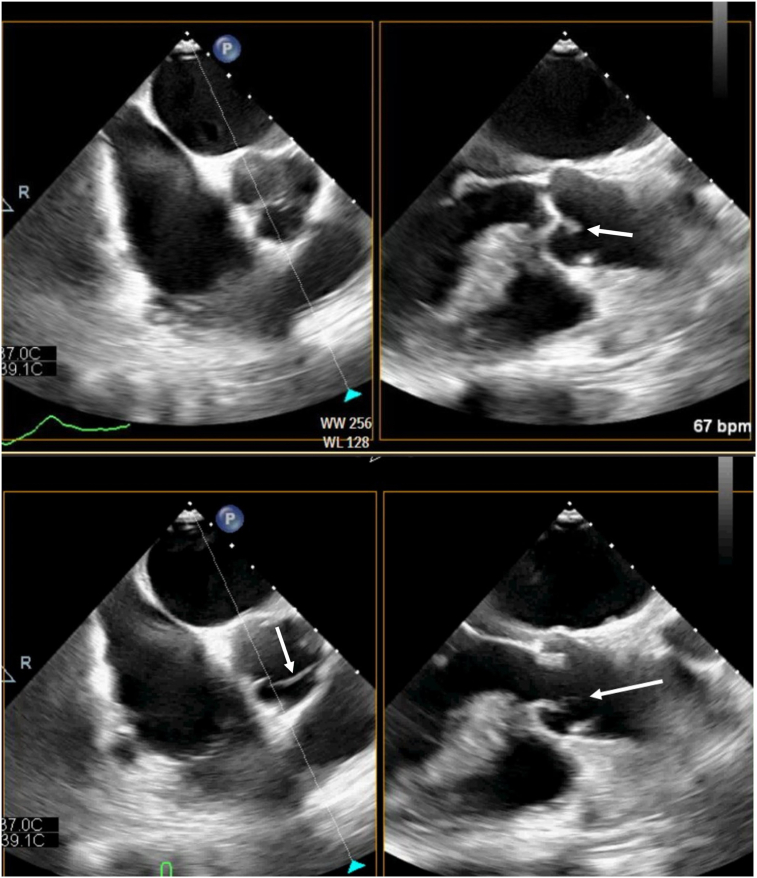 Figure 3
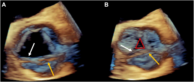 Figure 4
 Figure 5
Peer Reviews
No public reviews on file for this paper yet. If you reviewed it on a platform where reviews are public (OpenReview, ICLR, NeurIPS, ICML), you can paste yours below so the community can read it here.
Videos
No videos yet. Explain this paper in a talk, walkthrough, or lecture? Add one.
Taxonomy
TopicsCardiac Valve Diseases and Treatments · Infective Endocarditis Diagnosis and Management · Cardiac Structural Anomalies and Repair