Probabilistic Nested Model Selection in Pharmacokinetic Analysis of DCE-MRI Data in Animal Model of Cerebral Tumor
Hassan Bagher-Ebadian, Stephen L. Brown, Mohammad M. Ghassemi, Prabhu C. Acharya, Indrin J. Chetty, Benjamin Movsas, James R. Ewing, Kundan Thind

TL;DR
This paper introduces a new unsupervised method to improve pharmacokinetic analysis of DCE-MRI data in cerebral tumors by estimating the probability of different models per voxel.
Contribution
The novel contribution is the use of Kohonen-Self-Organizing-Map (K-SOM) to estimate probabilistic model contributions in DCE-MRI pharmacokinetic analysis.
Findings
The K-SOM probabilistic-NMS technique showed strong similarity to traditional NMS in identifying leaky tumor regions.
The new method produced microvasculature parameters less affected by arterial-input-function dispersion.
Estimated permeability parameters showed significant mean-percent-differences compared to traditional NMS.
Abstract
Best current practice in the analysis of dynamic contrast enhanced (DCE)-MRI is to employ a voxel-by-voxel model selection from a hierarchy of nested models. This nested model selection (NMS) assumes that the observed time-trace of contrast-agent (CA) concentration within a voxel, corresponds to a singular physiologically nested model. However, admixtures of different models may exist within a voxel’s CA time-trace. This study introduces an unsupervised feature engineering technique (Kohonen-Self-Organizing-Map (K-SOM)) to estimate the voxel-wise probability of each nested model. Sixty-six immune-compromised-RNU rats were implanted with human U-251N cancer cells, and DCE-MRI data were acquired from all the rat brains. The time-trace of change in the longitudinalrelaxivity ΔR1 for all animals’ brain voxels was calculated. DCE-MRI pharmacokinetic (PK) analysis was performed using NMS to…
Genes, proteins, chemicals, diseases, species, mutations and cell lines named across the full text — each resolved to its canonical identifier and authoritative record.
Click any figure to enlarge with its caption.
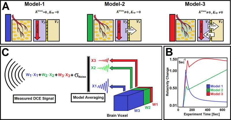 Figure 1
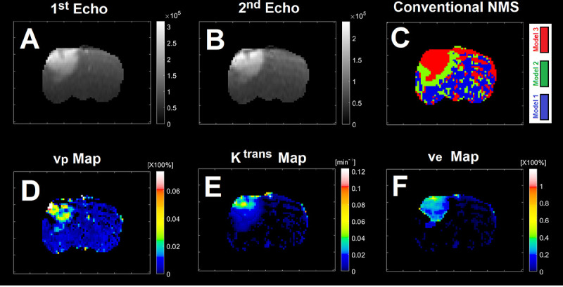 Figure 2
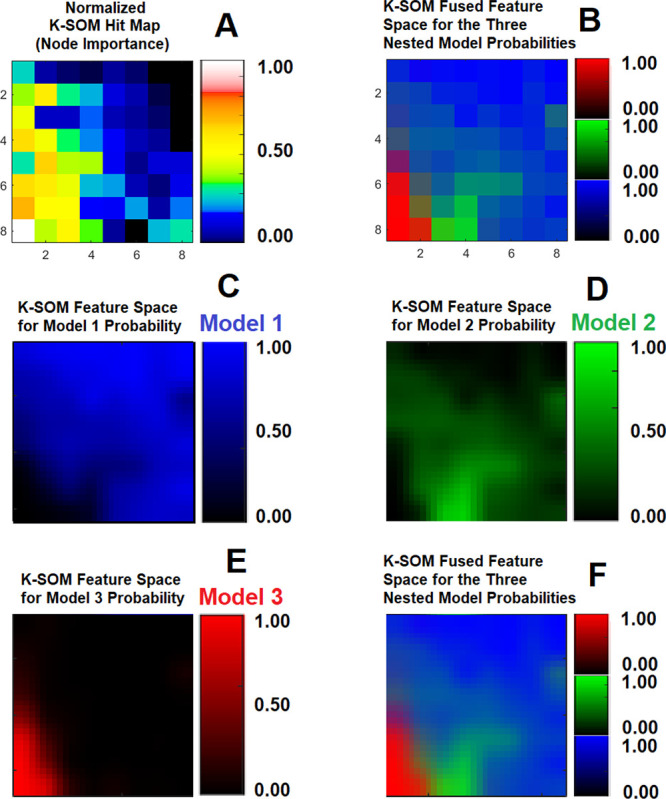 Figure 3
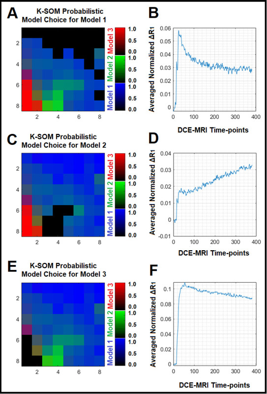 Figure 4
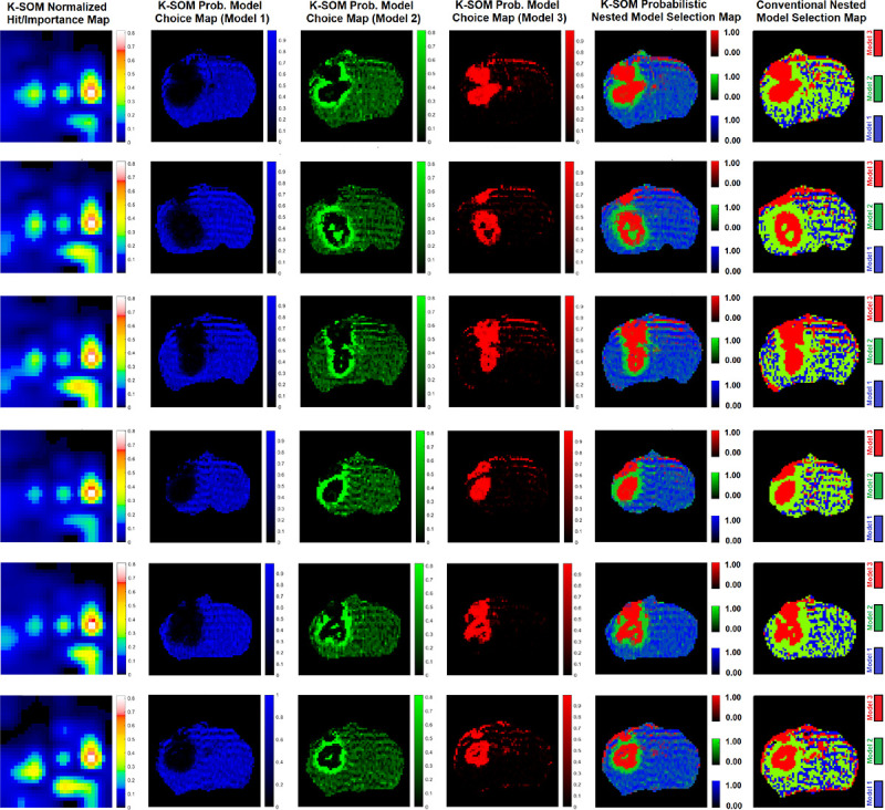 Figure 5
Peer Reviews
No public reviews on file for this paper yet. If you reviewed it on a platform where reviews are public (OpenReview, ICLR, NeurIPS, ICML), you can paste yours below so the community can read it here.
Videos
No videos yet. Explain this paper in a talk, walkthrough, or lecture? Add one.
Taxonomy
TopicsAdvanced MRI Techniques and Applications · Medical Imaging Techniques and Applications · MRI in cancer diagnosis