Intramural Longitudinal Sigmoid Fistula
Brecht Van Berkel, Vincent Sneyers, Geert Verswijvel

TL;DR
This paper discusses a rare complication of diverticulitis called a longitudinal intramural sigmoid fistula, which can be seen using CT scans.
Contribution
The paper highlights the identification and visualization of a rare complication of diverticulitis through CT imaging.
Findings
A longitudinal intramural fistula is a rare complication of diverticulitis.
Computed tomography (CT) can visualize this rare condition.
Abstract
Teaching point: A longitudinal intramural fistula is a rare complication of diverticulitis that can be visualised by computed tomography (CT).
Genes, proteins, chemicals, diseases, species, mutations and cell lines named across the full text — each resolved to its canonical identifier and authoritative record.
Click any figure to enlarge with its caption.
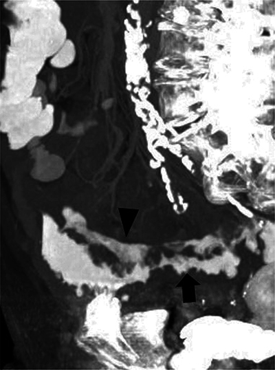 Figure 1
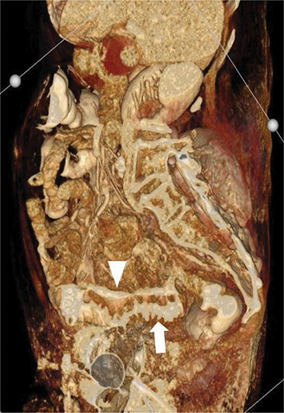 Figure 2
Peer Reviews
No public reviews on file for this paper yet. If you reviewed it on a platform where reviews are public (OpenReview, ICLR, NeurIPS, ICML), you can paste yours below so the community can read it here.
Videos
No videos yet. Explain this paper in a talk, walkthrough, or lecture? Add one.
Taxonomy
TopicsAnorectal Disease Treatments and Outcomes