Secondary Metabolites and Antioxidant Activity against Moko Disease as a Defense Mechanism of Musa spp. from the Ecuadorian Coast Area
Raluca A. Mihai, Vanessa A. Terán-Maza, Karen A. Portilla-Benalcazar, Lissette E. Ramos-Guaytarilla, María J. Vizuete-Cabezas, Erly J. Melo-Heras, Nelson S. Cubi-Insuaste, Rodica D. Catana

TL;DR
This study explores how banana plants from Ecuador's coast defend against Moko disease by producing antioxidant compounds.
Contribution
The study identifies specific secondary metabolites in Musa spp. that increase during Moko disease infection as part of the plant's defense mechanism.
Findings
Diseased Musa spp. samples showed higher accumulation of antioxidant secondary metabolites than healthy ones.
Over 40 compounds, including kaempferol and quercetin glycosides, were identified as part of the defense system against Ralstonia solanacearum.
LC-MS and antioxidant assays confirmed increased synthesis of active compounds during Moko disease symptoms.
Abstract
The Musa spp. represents the most commonly produced, transitioned, and consumed fruit around the globe, with several important applications in the biotechnology, pharmaceutical, and food industries. Moko disease is produced by Ralstonia solanacearum—a factor with a high impact on all crops in Ecuador, representing one of the biggest phytosanitary problems. Four of the most common varieties of Musa spp. were tested to identify the metabolic reaction of plants facing Moko disease. The phenolic and flavonoid content has been evaluated as a defense system, and the α-diphenyl-α-picrylhydrazyl free-radical-scavenging method (DPPH), free-radical-scavenging activity (ABTS), ferric-reducing antioxidant power (FRAP) assays, and liquid chromatography and mass spectrometry (LC-MS) have been adapted to analyze the active compounds with the antioxidant capacity necessary to counteract the pathogenic…
Genes, proteins, chemicals, diseases, species, mutations and cell lines named across the full text — each resolved to its canonical identifier and authoritative record.
Click any figure to enlarge with its caption.
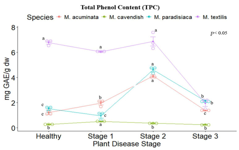 Figure 1
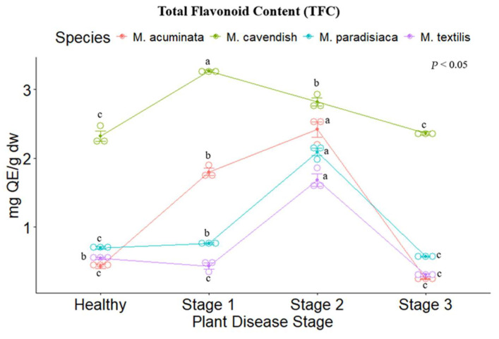 Figure 2
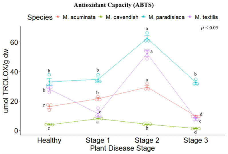 Figure 3
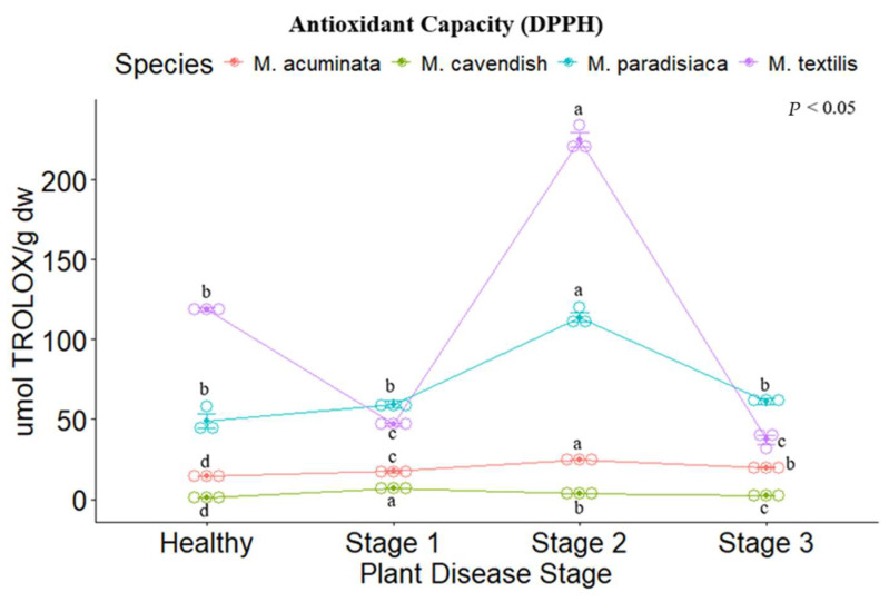 Figure 4
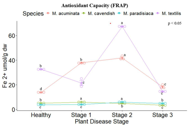 Figure 5
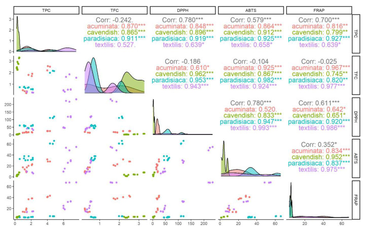 Figure 6
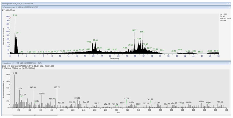 Figure 7
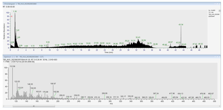 Figure 8
- —Universidad de Las Fuerzas Armadas-ESPE
- —Institute of Biology Bucharest, Romanian Academy
Peer Reviews
No public reviews on file for this paper yet. If you reviewed it on a platform where reviews are public (OpenReview, ICLR, NeurIPS, ICML), you can paste yours below so the community can read it here.
Videos
No videos yet. Explain this paper in a talk, walkthrough, or lecture? Add one.
Taxonomy
TopicsBanana Cultivation and Research · Plant Pathogenic Bacteria Studies · Cassava research and cyanide
1. Introduction
The Musa spp. (Musaceae family) enjoys increased interest since bananas represent the world’s most commonly produced, traded, and consumed fruit, contributing to food security and incomes, and being in fourth place in importance in numerous countries [1]. Ecuador is the largest exporter globally, among Africa, Asia, and Latin America, which produce and sell bananas [2]. Besides its economic importance, the interest in banana plants grew due to their numerous uses in pharmacy, biotechnology, and food [3].
Among the factors that may affect banana manufacture, diseases caused by bacteria have serious effects [4]; Ralstonia solanacearum is one of the top 10 bacterial pathogens worldwide [5], characterized by a wide host range and wide geographic distribution, making it one of the most destructive crop pathogens in the world [6]. An alert was issued in February 2023 regarding Moko disease in Ecuador, reported in Musa plantations in more than 12 provinces [7].
Moko disease represents one of the phytosanitary problems with a great economic impact on all banana varieties and plantain crops, which affects over 70% of yield losses [6], as reported in a variety of studies regarding the treatment of this disease. Over time, some studies have been concerned about the Moko disease in Musa spp. In this way, molecular approaches like RAPD (Random Amplified polymorphic DNA) and rep-PCR-based fingerprinting (Repetitive sequence-based Polymerase Chain Reaction [8], AFLP molecular markers (Amplified Fragment Length Polymorphism) [9], and real-time PCR [10] were used to investigate the genetic relationship between Ralstonia solanacearum and Musa spp. diseases. Different strategies were tested to control/or eradicate the Moko disease: resident varieties [11,12,13,14], banana leachates [15], insects and beneficial microorganisms [16].
R. solanacearum invades the vascular tissues, causing the leaf’s death, which reduces the ability to produce fruit. Insects and/or contaminated farming equipment instead of mechanical and soil are the principal transmission vectors [17]. Recent genetic studies showed that bacterial strains can survive more than 25 years, affecting native flora, organic matter in soils, and crop plants in tropical and subtropic zones [18]. Even though there are some good practices (chlorine dioxide) and resistance inducers in banana plants [19] for farmers to reduce the spread of the disease agent, no cure is available for Moko.
Secondary metabolites are directly involved in plant defense; factors like genetic, ontogenic, morphogenetic, and environmental factors may affect their biosynthesis/accumulation [20]. Recent studies have pointed out that banana flavonoids can form complex compounds with extracellular proteins that can damage the cell membrane of bacteria, followed by the release of intracellular compounds [21]. Our study aimed to demonstrate that the antioxidant capacity resulting from the biosynthesis of secondary metabolites may act as a defense mechanism against the Moko attack in Musa spp. To achieve this aim, four of the most commonly used Musa spp. were investigated in our research, such as Musa cavendish Paxton [22]—the most widespread edible variety [23], Musa paradisiaca L.—a plantain used as a nutritional and therapeutic source [24], Musa textilis Née, an important source of fibers [25] and Musa acuminata Colla—the most frequent species [26].
2. Materials and Methods
2.1. Chemicals
To quantify the different secondary metabolites, the following chemicals (Sigma-Aldrich Chemical Co.—St. Louis, MO, USA) were used: 2.2—diphenyl—1—picrylhydrazyl, diammonium salt of 2.2—azinobis (3–ethylbenzothiazoline– 6-sulfonic acid), 2.4.6-tri(2-pyridyl)-triazine, iron (III) chloride hexahydrate and Folin–Ciocalteu reagent, 6-hydroxy-2.5.7.8-tetramethyl chroman-2-carboxylic acid, 3.4.5-trihydroxy benzoic acid, potassium persulfate, iron (III) sulfate heptahydrate, quercetin, and ethanol. All the reagents were purchased from commercial providers.
2.2. Sample Collection and Processing
This study was carried out with plants of the genus Musa spp., from farms located in Santo Domingo de los Tsáchilas province (0°05′01.6″ N, 79°26′32.8″ W), Canton La Concordia, km 38 road Santo Domingo—Quinindé. Healthy plants and plants with Moko disease symptomatology, at different stages of disease development, were collected. Each degree of infection (disease score) has been characterized and identified based on the following characteristics implemented by the Government of Kerala in 2014: Stage 1 (initial stage) symptoms are characterized by the yellowing of the central leaf, transitioning from yellow to green, weakening, and eventually breaking at the junction with the petiole. As the infection progresses, it leads to wilting and drying of young leaves, spreading to older ones. Older leaves then exhibit yellow bands with dark margins on their edges. In Stage 2 (intermediate stage), symptoms appear swiftly in the suckers, characterized by the progressive yellowing and wilting of older leaves. Additionally, small suckers exhibit a gradual death of the central leaf, which extends toward the outer leaves. In Stage 3 (advanced stage), symptoms in infected corms become evident through transverse cuts, revealing brown or black bands in the vascular bundles infected by the bacteria. Diseased pseudostems excrete bacterial exudate, while internally, vascular bundles exhibit a light to dark brown color due to blockage by extracellular polymeric substances. Plants without clusters display symptoms characterized by the grouping of vascular bundles near the pseudostem center, with significant blockages primarily observed in the petioles of the initially infected leaves, becoming less apparent toward the periphery. All the collected banana plants were in the vegetative stage, young plants of approximately 1 year old. The samples were transferred in a cooler to the university laboratory in Cantón Rumiñahui.
2.3. Active Ingredients Extraction
To determine the relationship between the antioxidant character and the phytochemical compounds involved in the defense of plants against Moko disease, in the first phase, the impurities were removed from the leaves by washing them with distilled water, and afterwards, the leaves were dried and ground (5 g) and placed in 25 mL of ethanol of HPLC (High Performance Liquid Chromatography) grade 99.5 (v/v), overnight, in the dark, at 4 °C.
2.4. Active Ingredients Determination
The Folin–Ciocalteu method was used to determine the phenolic concentration in the banana leaves [27]. Solutions from the ethanolic extracts (0.4 mL), Folin–Ciocalteu reagent (2 mL of 10% (v/v), and 7.5% Na_2_CO_3_ (1.6 mL) were incubated at room temperature for 30 min. The 765 nm absorbance was used to measure the concentrations. Gallic acid (0–250 mg/L concentration) was used for the calibration curve (y = 0.0112x + 0.1759, R^2^ = 0.9794).
A colorimetric method using aluminum chloride described by Pekal et al. [28] was used for the determination of the flavonoid content. Quercetin was used for the realization of the standard solution. The standard calibration curve was performed at a concentration between 0 and 1.5 mg L^−1^, obtaining the equation y = 1.4566x + 0.0265 with a correlation factor R^2^ = 0.9935. To the crude extracts of the plant sample (1 mL) was added 1.5 mL of the solvent, 100 µL of CH_3_COONa (1 M), 100 µL of AlCl_3_ (10% v/v), and distilled water (2.3 mL). The samples are covered for 40 min at room temperature. In the blank sample, the aluminum chloride was not placed. Finally, the samples were measured at 435 nm absorbance.
2.5. Antioxidant Capacity Determination
A protocol established by Sachett et al. (2021) [29] and modified by Thaweesang [27] was used for the determination of the antioxidant capacity through the 2,2-diphenyl-1-picrylhydrazyl (DPPH) radical. A stock solution of 1 µg L^−1^ was produced; 2 mL of stock was added to every laboratory test tube, including the 0.1 mL crude extracts sample. After the test tubes were left to incubate for 30 min at room temperature, the absorbance was measured at 517 nm and the radical scavenging activity was calculated using the following formula: control absorbance—sample absorbance/control absorbance ×100. The calibration curve (y = 158.07x − 1.6766, R^2^ = 0.9955) was produced using a Trolox solution (positive control) with a concentration between 0 and 0.625 mM.
The establishment of the antioxidant capacity by 2,2′-azino-bis(3-ethylbenzothiazoline-6-sulfonate) radical cation (ABTS^*+^) free radical scavenging (obtained from the reaction between ABTS (7 mM) and potassium persulfate) was based on a study by Kuskoski et al. [30]. After incubating the solution for 12–24 h, it was diluted in absolute ethanol until 0.7 ± 0.1 at 754 nm absorbance was obtained. The inhibitory capacity was determined after the reaction between the ABTS solution (2 mL) and sample (20 µL) for 7 min at room temperature and in darkness; absolute ethanol was used for the blank. The calibration curve was established between a 0 and 2.5 mM concentration, obtaining the following equation y = 31.995x + 3.9568 with a correlation factor R^2^ = 0.9697, using Trolox (like a positive control).
To determine the antioxidant capacity based on the Ferric ion Reducing Antioxidant Power (FRAP) test, a FRAP solution was prepared from acetate buffer 300 mM (100 mL) pH 3.6 + HCl (40 mM) + FeCl_3_ · 6 H_2_O (20 mM) in the proportion of 100: 10: 10 according to the method of Rajurkar et al. (2011) [30]. The absorbance of the mixture was read at 593 nm. The standard curve (y = 0.3654x − 0.0532, R^2^ = 0.9686) was performed by taking different concentrations of FeSO_4_ · 7H_2_O ranging from 0 to 5 mM. The solution consisting of sample (100 µL), distilled water (300 µL), and FRAP solution (3 mL) previously prepared was covered for half an hour at room temperature. The FRAP value (mmol Fe^2+^ g sample) was obtained by the following formula:
2.6. LC-MS Determination
High-resolution LC-MS was used to determine the bioactive compounds in the samples of stem and leaves from M. cavendish at stage 1 of Moko disease. The plant extractions were established using a modified method based on Tohma et al. [31]. Ethanolic extracts were obtained from lyophilized samples represented by leaf and stem (1 g) and 80% ethanol (20 mL) maintained for 2 h at 30 °C and centrifuged for 10 min at 5000 rpm at 4 °C. Afterwards, the extracts were filtered and a rotary evaporator at 30 °C was used for ethanol removal. The samples were kept in well-covered plastic tubes until analysis was performed at −20 °C.
Identification and detection of different metabolites were performed by HPLC equipment (Vanquish Thermo Fisher Scientific (Waltham, MA, USA)) and Mass Detector (Ion Trap Thermo Fisher Scientific (Waltham, MA, USA)). The samples were eluted through an Accucore Vanquish 150 × 2.1 mm column at 35 °C and a 0.5 mL/min flow rate [32]. A 10 μL volume of 0.1% formic acid was used as a mobile phase to inject into the HPLC. MS scanning was used to identify the compounds from the samples based on the retention time comparison of each peak and the monitoring of the ion pairs in a standard solution [33]. The compounds were identified by comparing them with those of the standard compounds available in databases (PubChem, ChEBI, Metlin, HPLC, and the literature data) using the MZmine 2.53 software [34].
2.7. Statistical Test
The RStudio statistical program (1.3.8 version) was used. Significant differences were analyzed through a two-way ANOVA test (p < 0.05). All the experiments were realized in triplicate, with the values being presented as the mean ± SD. The correlation between the secondary metabolites analyzed and the antioxidant power was evaluated by Pearson’s correlation coefficient, while the correlation matrix was depicted as a scatter plot matrix.
3. Results
3.1. Active Ingredient Determination
The total phenolic content was evaluated for each disease stage per species. For M. cavendish, the highest phenolic content was registered in Stage I (0.507 ± 0.025 mg GAE g dw), Stage II (0.358 ± 0.052 mg GAE g dw), followed by healthy plants (0.27 ± 0.036 mg GAE g dw), while Stage III presented the lowest values. In contrast, for M. acuminata, M. paradisiaca and M. textilis, the highest phenolic content was registered in Stage II (4.114 ± 0.145 mg GAE g dw; 4.604 ± 0.215 mg GAE g dw, and 6.868 ± 0.526 mg GAE g dw, respectively), while the other stages presented a similar tendency. M. textilis showed the highest phenolic content of all the evaluated Musa spp. (Figure 1).
M. cavendish showed the highest flavonoid concentration in contrast to the other species, with Stage I presenting the highest value (3.266 ± 0.0295 mg QE/g dw). The other Musa spp. show a similar trend to the phenolic content, as the highest value was reported for M. acuminata, M. paradisiaca, and M. textilis, appearing for Stage II (2.419 ± 0.197, 2.091 ± 0.0937, 1.682 ± 0.154, respectively), followed by the healthy stage, and Stages I and III with similar values. The lowest flavonoid content was reported for M. textilis (Figure 2).
3.2. Antioxidant Activity Determination
The antioxidant capacity of the four Musa spp. was evaluated through ABTS, DPPH, and FRAP assays. Significant differences were found between the disease stages for each species, following the same trend as their respective results concerning the secondary metabolites analyzed. For the ABTS and DPPH methods, M. paradisiaca (ABTS: 62.545 ± 2.524 µmol TEAC (Trolox Equivalent Antioxidant Capacity) g dw, DPPH: 113.997 ± 4.451 µmol TEAC g dw) and M. textilis (ABTS: 52.212 ± 2.742 µmol TEAC g dw, DPPH: 225.212 ± 6.708 µmol TEAC g dw) showed the highest values for Stage II. (Figure 3 and Figure 4), while for the FRAP assay. M. acuminata (41.803 ± 1.179 µmol Fe ^2+^ g dw) and M. textilis (67.24 ± 0.284 µmol Fe ^2+^ g dw) presented the highest values (Figure 5).
The correlation between the two analyzed secondary metabolites was raised for the four Musa spp. The contents were correlated with the three methods of antioxidant capacity evaluation used, with each plant showing a higher positive correspondence (R > 0.90) for the total phenolic content and flavonoid content. In contrast, for the phenolic content. M. textilis showed a lower correlation, albeit still positive with the ABTS (R = 0.64), DPPH (R = 0.66), and FRAP (R = 0.64) assays (Figure 6).
3.3. LC-MS Determination
LC-MS was used to determine the bioactive compounds in the samples of stem and leaves from M. cavendish at Stage 1 of Moko disease. The compounds in the extracts were identified based on their molecular mass and retention time. The phenolic extracts from the stem (34 identified compounds) and leaf (61 identified compounds) samples yielded similar HPLC profiles. However, differences were observed between both samples in terms of the identified phenolic compounds. The leaf and stem samples showed a similar content of phenol precursors such as shikimic acid, caffeoyl alcohol, coumaroyl, and derivatives of amino acids. Flavonoids were exclusively found in the leaf samples, with derivatives of quercetin (spiraeoside, rutin), kaempferol (astragalin, kaempferol-7-O-neohesperidoside), naringenin derivatives, and other flavonoid glycosides. Flavones such as isoorientin and scutellarin 4′-methyl ether had also been identified in the leaf samples. Citric acid, gibberellic acid, and terpenes (e.g., 7-O-methyl rosmanol) were identified mostly in the stem (Table 1, Figure A1 and Figure A2).
4. Discussion
Banana, as a key crop for food security, is considered the primary food for several developing countries [13]. However, plants from the genus Musa spp. are susceptible to some pathogens, including R. solanacearum race 2. The wilt disease is determined by bacterial accumulation, which blocks the vessels, stopping the sap flow [43]. Bacterial wilt lowers the yield in banana production [13]; therefore, treatment and management of this bacterial disease involve the achievement of advanced technologies together with research. The understanding of the interaction between Musa spp. and pathogens will allow for identifying new strategies to be used to control the diseases without damaging the environment. Physical barriers, vulnerability, and phytoalexin yielding are some of the recognized traits used by Musa plants as defense mechanisms against pathogens [44,45]. Although the crops are indeed the most affected, plants possess different mechanisms to protect themselves, such as the development of secondary metabolites to eliminate the pathogen from its system.
In our case, all four of the most commonly used Musa spp. investigated (M. cavendish, M. paradisiaca, M. textilis, and M. acuminata) showed a similar trend in front of infection with R. solanacearum race 2. The diseased samples exhibited a higher antioxidant activity compared to the healthy samples, and we attempted to show that this is attributed to the presence of secondary metabolites (phenolic compounds) through the LC-MS analysis to reinforce the idea that secondary metabolites and implicitly antioxidant activity represent a defense mechanism against the pathogen that causes Moko disease. Plants possess physical and chemical barriers involved in their defense; they have a strategy to help their survival when in contact with some biotic or abiotic stresses, implicating the synthesis of secondary metabolites, which is well known as part of the plant immune system. A strong emphasis is placed on antioxidant activity to increase plant protection against the pathogen. The synthesis of metabolites comes from the primary metabolism (glycolysis, Krebs cycle, or shikimate pathway) that depends on the degree of stress to which the plant is subjected and can trigger variance in the levels of secondary metabolites, some of them toxic when stored in plant cells [46]. Synthesized phenolic compounds like simple phenols, flavonols, dihydrochals, and cones phenolic acids are antibiotic compounds that generate a response against pathogens. Phytoalexins, synthesized de novo, manage to inhibit a diversity of microorganisms by their accumulation at the infected site [47].
The antioxidant capacity of plants is related to the defense of both types of antioxidants (enzymatic and non-enzymatic) to escape from the toxic effects of free radicals. Their genetic configuration confers a great capacity to synthesize secondary metabolites under biotic or abiotic stress. Some compounds act like substrates in enzyme-catalyzed detoxification reactions and have central and interrelated functions [48,49,50].
Likewise, there is a considerable difference between plant developmental stages. The vegetative stage of the samples shows a lower content of secondary metabolites and antioxidant activity. The bioactive compounds produced depend on the environmental conditions to generate an adequate impact on the development of the metabolic pathways associated with their biosynthesis [51]. In the vegetative state, plants use photosynthesis and carbon assimilation for growth, development, and defense, and when exposed to some stress, can alter the storage and synthesis of metabolites. These include the harvest time, exposure to factors such as light, temperature, osmotic potential, nutrition, growth regulators, biotic inducers, and fruit-ripening stage, among others [52].
Based on their elemental role in plant protection against different agents, phenolics are known as antioxidants [53]. Phenolic compounds exhibiting significant antioxidant compounds were identified in each Musa spp. The predominant polyphenols in the plant defense mechanism are flavonoids, which may be classified as flavonols, isoflavonols, flavones, flavanones, catechins, and anthocyanidins [54]. New research has shown the involvement of flavonoids in plant protection, playing a significant role in the neutralization of free radicals [55], especially in terms of the highly sensitive antimicrobial effect on pathogens, in which compounds such as naringenin, kaempferol, quercetin, and dihydroquercetin stand out [56]. Recent studies have described the antibacterial effect of phenolic compounds obtained from plantain leaves against some Gram-negative bacteria (Escherichia coli, Staphylococcus, Pseudomonas) species [57,58]. The bacterium Ralstonia solanacearum race 2 (Smith, 1896) is a Gram-negative bacillus with high genetic variability that affects the vascular system of the plant [59]. The phenolic compounds identified in the current study have shown antibacterial activity toward the infection since this strain is responsible for causing the Moko disease in Musa spp., whose direct effect is aggravated by the capacity of its causative agent to remain in the soil for a long time, disabling the immediate replanting of the affected lots [60], highlighting the importance of describing plant defense mechanisms to reach a better understanding of the infection. In our current study, these compounds and their derivatives have been identified, from kaempferol (astragalin, kaempferol-7-O-neohesperidoside) and quercetin glycosides (spiraeoside, rutin), as well as flavones like isoorientin and scutellarein 4′-methyl. Quercetin and its derivatives also play significant roles in plant protection from the effects of UV radiation and/or osmotic stress, in which glycosylated derivatives are involved in osmoregulation [61]. In addition, flavonoids are known for their antioxidant capacity by decreasing ROS levels by inhibiting prooxidant enzymes, cyclooxygenase, and lipoxygenase [62].
Furthermore, signaling molecules have been identified; products of oxidative processes’ ROS (7-methyl-rosmanol), supporting the idea of the loss of some acids (carnosic acid) with antibacterial activity against both bacteria types (Gram-positive and Gram-negative) causing the accumulation of oxidized derivatives under oxidative degradation by ROS [63,64]. Microbial attack and the oxidative state of plants mediate the activation of the plant protection mechanisms against stress through different signaling pathways, which conduct the production of various protein and non-protein compounds with roles in protection [65], such as salicylic acid (2-hydroxyhippuric acid), which has also been identified in our samples through LC-MS; salicylic acid levels are known to increase during different type of infections (viruses, fungi, insects, bacteria), while exogenous treatment with salicylic acid improves the protection system of the host [66].
Bananas and plantains are the most commonly consumed as food and are used in medicine around the world, being an attractive source of bioactive compounds. These compounds with desirable biological properties for humans are also implicated in the plant protection strategy against pathogens, as in the case of phenolics, which are a key aspect in Musa spp. in the protection mechanism, as it has been found in the present study. A higher content of phenols, flavonoids, and antioxidant capacity was noted in the late stages of the infected samples compared to healthy samples, which showed a lower amount of phenolics and activity.
Identification of the phenolic compounds demonstrated the presence of relevant flavonoids that are involved in the defense mechanism of plants and which are known for their antibacterial activity related to infection by Gram-negative bacteria like R. solanacearum responsible for the Moko disease affecting the Musa spp. genus.
5. Conclusions
Bananas and plantains belonging to the genus Musa are largely consumed all over the world as food staples and for medicinal purposes, being an interesting source of bioactive secondary metabolites. These compounds, with valuable biological properties for humans as antioxidants, also play a pivotal role in the plant’s defense mechanism against pathogens. For example, phenolic compounds are essential to the defense strategy of Musa spp., as demonstrated in the current study, enhancing the antioxidant profile of these plants. Infected samples showcased a higher concentration of phenols, flavonoids, and antioxidant activity compared to healthy ones, which exhibited lower levels of phenolics and activity. The analysis of the phenolic compounds revealed the presence of significant flavonoids that participate in plant defense mechanisms. The flavonoids, including kaempferol, quercetin, and their glycosides, which we found in the banana samples, have antibacterial properties implicated in the fight against various pathogenic bacteria by interfering with their growth and survival.
The reference list from the paper itself. Each links out to its DOI / PubMed record.
- 1International Network for the Improvement of Banana and Plantain, International Plant Genetic Resources Institute 2000 Available online: https://cgspace.cgiar.org/handle/10568/105424?show=full(accessed on 15 June 2023)
- 2FAO Banana Market Review—Preliminary Results 2022 FAO Rome, Italy 2022
- 3Lopes S. Vanz Borges C. de Sousa Cardoso S.M. de Almeida Pereira da Rocha M.F. Maraschin M. Banana (Musa spp.) as a Source of Bioactive Compounds for Health Promotion Handbook of Banana Production, Postharvest Science, Processing Technology, and Nutrition 1st ed. Siddiq M. Ahmed J. Lobo M.G. John Wiley & Sons Ltd.Hoboken, NJ, USA 2020
- 4Blomme G. Dita M. Jacobsen K.S. Pérez V.L. Molina A. Ocimati W. Poussier S. Prior P. Bacterial Diseases of Bananas and Enset: Current State of Knowledge and Integrated Approaches Toward Sustainable Management Front. Plant Sci.201720129010.3389/fpls.2017.0129028785275 PMC 5517453 · doi ↗ · pubmed ↗
- 5Mansfield J. Genin S. Magori S. Citovsky V. Sriariyanum M. Ronald P. Dow M. Verdier V. Beer S.V. Machado M.A. Top 10 plant pathogenic bacteria in molecular plant pathology Mol. Plant Pathol.20121361462910.1111/j.1364-3703.2012.00804.x 22672649 PMC 6638704 · doi ↗ · pubmed ↗
- 6Denny T. Plant pathogenic Ralstonia species Plant-Associated Bacteria Gnanamanickam S.S. Springer Netherlands Cham, The Netherlands 2006573644
- 7Banana Moko: A Silent Threat 2022 Available online: https://www.ecuadortimes.net/banana-moko-a-silent-threat/(accessed on 15 June 2023)
- 8Thwaites M. Eden-Green S. RAPD and rep PCR-based fingerprinting of vascular bacterial pathogens of Musa spp.Plant Pathol.19994812112810.1046/j.1365-3059.1999.00321.x · doi ↗