Analysis of CYP1B1 Polymorphisms in Lung Cancer Patients Using Novel, Quick and Easy Methods Based on CAPS and ACRS-PCR Techniques
Adam Dąbrowski, Maciej Nowicki, Aleksandra Budzyńska, Jakub Suchodolski, Rafał Ogórek, Mariusz Chabowski, Katarzyna Przywara

TL;DR
This study introduces cost-effective methods to identify CYP1B1 gene polymorphisms in lung cancer patients, finding higher frequencies in this group compared to the general European population.
Contribution
The paper presents novel, low-cost CAPS and ACRS-PCR methods for unambiguous identification of four CYP1B1 SNPs without the need for sequencing.
Findings
The developed methods enable specific identification of four CYP1B1 polymorphisms with no need for sequencing.
Lung cancer patients showed 1.5 to 2.1 times higher frequencies of the analyzed SNPs compared to the European population.
The methods reduce research time, cost, and error rates using basic lab equipment and careful primer/enzyme design.
Abstract
Within the sequence of the CYP1B1 gene, more than 50 polymorphisms, resulting from single-nucleotide polymorphisms (SNPs), have been described. Some of them play an important role as specific genetic markers in the process of carcinogenesis and for therapeutic purposes. In this publication, we present methods we have developed that enable the specific and unambiguous identification of four polymorphisms that result in amino acid changes: c. 142C > G, c. 355G > T, c. 1294C > G, and c. 1358A > G. Our studies are based on cleaved amplified polymorphic sequences (CAPSs) and artificially created restriction site (ACRS) PCR techniques; therefore, they require only basic laboratory equipment and low financial outlays. Utilizing the described methods allows for the reduction of research time and cost, and the minimization of errors. Their effectiveness and efficiency depend on the careful…
Genes, proteins, chemicals, diseases, species, mutations and cell lines named across the full text — each resolved to its canonical identifier and authoritative record.
Click any figure to enlarge with its caption.
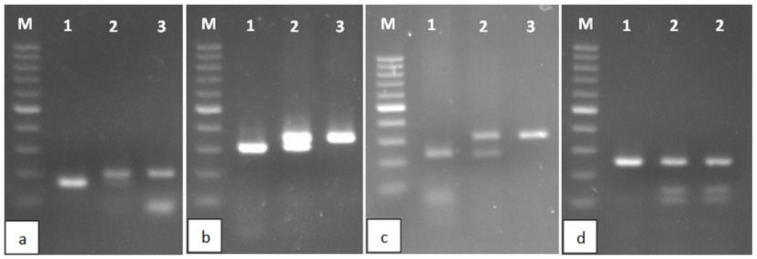 Figure 1
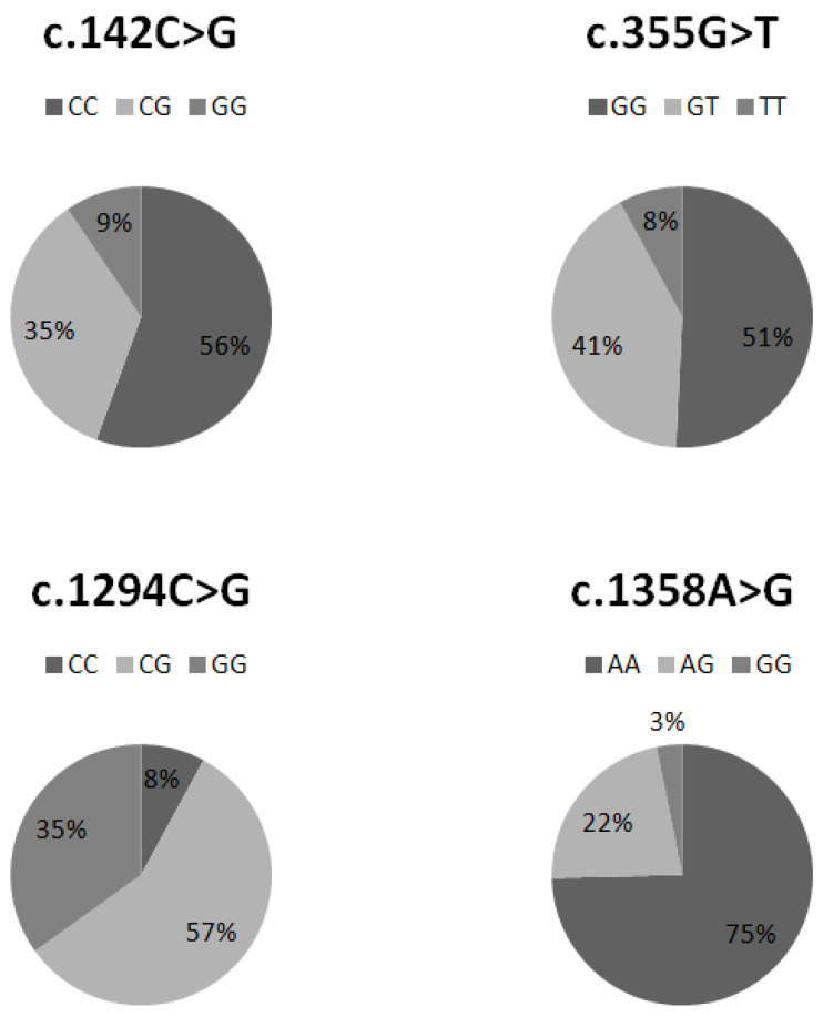 Figure 2
Peer Reviews
No public reviews on file for this paper yet. If you reviewed it on a platform where reviews are public (OpenReview, ICLR, NeurIPS, ICML), you can paste yours below so the community can read it here.
Videos
No videos yet. Explain this paper in a talk, walkthrough, or lecture? Add one.
Taxonomy
TopicsMetabolomics and Mass Spectrometry Studies · Carcinogens and Genotoxicity Assessment · Pharmacogenetics and Drug Metabolism
1. Introduction
Cytochrome P450 (CYP450) is found in every class of living organism, including Archaea, and as organisms have evolved, its functionality has increased [1]. Its main role is detoxification, and in the human body, it occurs in almost every cell type, but it achieves the greatest activity in the cells of the small intestine, liver, and adrenal medulla [2]. CYP450 enzymes are characterized by low substrate specificity. They participate in hydroxylation, e.g., steroid hormones, bile acids, prostaglandins, or xenobiotics, including drugs, pesticides, tobacco smoke, and food additives [3]. In terms of oncology, a very interesting member of the CYP450 superfamily is the enzyme CYP1B1, which has been placed in a list of cancer antigen prioritization for targeted therapies by the National Cancer Institute [4]. The CYP1B1 gene is located on chromosome 2p22.2 and consists of 12,000 base pairs divided into three exons and two introns. The mature transcript contains over 5000 base pairs and encodes a protein of 543 amino acids [5]. Unlike most CYPs, its expression is low in normal tissues; however, it is often overexpressed in cancer [6]. Like all cytochromes P450, it catalyzes oxidation reactions. Its main substrates are 17β-estradiol and polycyclic aromatic hydrocarbons (PAHs), which become carcinogens and can cause double-stranded DNA breaks [6,7]. Within the sequence of the CYP1B1 gene, more than 50 polymorphisms resulting from SNPs have been described [8]. The best-known polymorphisms that result in amino acid changes include c. 142C > G (R48G), c. 355G > T (A119S), c. 1294C > G (L432V), c. 1358A > G (N453S) [7]. There are many scientific reports indicating a link between the presence of CYP1B1 polymorphisms and the risk of lung cancer. They focus primarily on the c. 1294C > G, c. 1358A > G, and c. 355G > T isoforms [9,10,11,12,13,14,15]. Unfortunately, due to heterogeneous groups, the results are not unanimous.
CYP1B1 polymorphisms play an important role as specific genetic markers in the process of carcinogenesis and for therapeutic purposes. Therefore, in this publication, we have developed fast, specific, and cost-effective methods for identifying c. 142C > G, c. 355G > T, c. 1294C > G, and c. 1358A > G polymorphisms using CAPS and ACRS-PCR techniques. In addition, using these methods, we examined 63 patients diagnosed with lung cancer. We subjected the obtained results to statistical analysis, considering histopathological diagnosis and cancer stage assessment.
2. Results
2.1. Diagnosis of Polymorphism in the CYP1B1 Gene
2.1.1. c. 142C > G
In the case of this polymorphism, we observe a substitution of cytosine base for guanine [C/G]. In order to amplify the appropriate DNA fragment, appropriately constructed primers were used that changed the amplicon fragment beyond the mutation locus (Table 1). Consequently, the use of the restriction enzyme HpaII resulted in the cleavage of the wild-type gene fragment at only one site, yielding 171 and 33 bp products. In contrast, the allele with the mutation remained undigested (204 bp) (Table 2, Figure 1).
2.1.2. c. 355G > T
The CYP1B1 c. 355G > T polymorphism was identified by digesting the amplified 364 bp gene fragment with NaeI restriction enzyme. This endonuclease recognizes a sequence at a potential mutation site. Therefore, strand scission occurred only in the wild type, producing 290 and 74 bp products. The mutant remained uncut (364 bp) (Table 2, Figure 1).
2.1.3. c. 1294C > G
The amplified 310 bp gene fragment was subjected to the restriction enzyme OliI, which recognizes sequences characteristic of the wild form of the gene. As a result, in the case of the L423 form, two products were obtained: 228 and 82 bp. In the presence of the c.1294C > G mutation, the product was not digested and remained at 310 bp (Table 2, Figure 1).
2.1.4. c. 1358A > G
After amplification, a product of 324 bp was obtained. The MwoI enzyme recognizes the 5′-GCNNNNNNNGC-3′ sequence. In the amplified fragment of the wild form of the gene, this sequence is present in two places, while in the mutated form, there is an additional place within the mutation itself. Thus, treatment of the amplicone with the MwoI enzyme resulted in products of 36, 35, and 253 bp in the case of the wild-type allele and 36, 35, 108, and 145 bp in the presence of a nucleotide change (Table 2).
Overall information on the histopathological diagnosis, tumor staging, gender, age, smoking status, and genotypes of individual polymorphisms of all 63 examined patients is attached as Table S1 (Supplementary Materials).
2.2. Frequency of Occurrence of Individual Polymorphisms in the Analyzed Population of Lung Cancer Patients
The percentage distribution of individual genotypes in the c. 142C > G, c. 355G > T, 1294C > G, and c. 1358A > G polymorphisms is illustrated in Figure 2.
2.3. Statistical Analysis between the Presence of SNPs and the Type and Stage of Cancer
During the study, we observed that the identified SNPs did not correlate with either the type or progression of the tumor disease. Each SNP was statistically insignificant when using the χ^2^ test at α = 0.01 (Table S2, Supplementary Materials). Thus, we conclude that neither SNP nor the size of the primary tumor, involvement of lymph nodes, or distant metastasis (TNM staging) affects the development of specific tumor types (glandular, squamous, or small-cell carcinoma).
3. Discussion
Lung cancer ranks first in terms of morbidity and mortality from malignant tumors in the world. It is the second most commonly diagnosed cancer in men following prostate cancer, and in women following breast cancer [16]. In 2018, the Global Cancer Observatory (GLOBOCAN) recorded over 2 million new cases of lung cancer and 1.8 million deaths caused by it [16]. Such results are influenced, among others, by the increase in urbanization, environmental pollution (mainly by PAHs), but, above all, by active and passive smoking [17]. Benzo[a]pyrene, the main component of tobacco smoke and environmental pollutants, is activated with the use of cytochrome P450 enzymes [18]. The main role in this process is played by the CYP1A1 and CYP1B1 enzymes, which convert PAHs into an intermediate form—epoxides—which are then subjected to the action of epoxide hydrolase, which modifies them into reactive mutagens [18]. The influence of polymorphisms in the CYP1B1 gene in this process is not fully understood, but it is suggested that they increase the process of activation of mutagenic compounds [7].
The mutations we study are directly related to the formation of cancers conditioned by the influence of environmental factors, such as smoking, environmental pollution from combustion products of low-quality coal, petroleum hydrocarbons and plastics, contact with organic solvents, and the use of steroid hormones. The methods used in the study enable the determination of c. 142C > G, c. 355G > T, c. 1294C > G, and c. 1358A > G polymorphisms in the CYP1B1 gene. They are based on the amplification of CYP1B1 gene fragments and their digestion with appropriate restriction enzymes. The products are visualized using standard horizontal electrophoresis, allowing for unambiguous test result determination. Therefore, each of the diagnostic stages requires only basic laboratory equipment and low financial outlays. This way of obtaining results does not require further confirmation by sequencing the DNA of each patient [19]. The effectiveness and efficiency of the research presented by us is based on the careful design of appropriate primers and the precise selection of suitable restriction enzymes. Some of our restricted products are less than 40 bp in size (Table 2); therefore, they may appear faint in typical agarose gel. However, it should be noted that the rest of the restriction cleavage products from the same amplicon are sufficiently large and apparently differentiated depending on the genotype. Thus, the difficulty in reading small bands does not affect the diagnostic effectiveness.
Germline mutations are the basis of hereditary lung cancers [20]. The CYP1B1 gene polymorphism can be homozygous or heterozygous. The presented tests allow us to determine whether a given patient has a mutation in one or two copies of the gene. This information may impact the course of the disease or treatment decisions. Studies suggest that patients who carry both mutated copies of the gene are characterized by more severe symptoms and worse response to treatment then heterozygotes [20]. Interestingly, the isoforms of this enzyme may also have a pharmacogenetic aspect. It has been observed that CYP1B1 isoforms can determine the effectiveness of treatment with, among others, taxanes or daunorubicin [21,22,23,24]. It must be noted that CYP1B1 does not metabolize these drugs.
According to the National Institutes of Health, the frequency of SNPs resulting in the c. 142C > G (rs10012), c. 355G > T (rs1056827), c. 1294C > G (rs1056836), and c. 1358A > G (rs1800440) polymorphisms in the European population is ~24, ~29, ~43, and ~17%, respectively. In our study group of 63 patients with lung cancer, we noted a higher frequency of the analyzed mutations, equal to approximately 44, 49, 92, and 25%, which corresponds to an increase of approximately 1.8, 1.7, 2.1, and 1.5 times, respectively. The most significant increase was observed in the c. 1294C > G polymorphism. This observation coincides with the conclusion presented in a meta-analysis from 2014, which showed a correlation between the occurrence of the L432V polymorphism and the development of lung cancer in the Caucasian, Asian, and African American populations [25]. It has been observed that individuals with the wild-type genotype (CC) exhibit higher levels of CYP1B1 gene expression compared to GG homozygotes [26]. Exposure to tobacco smoke or environmental pollution implies that individuals with the mutated genotype are unable to effectively remove toxins from cells, contributing to DNA damage and the accumulation of other mutations [26].
In conclusion, the methods presented by us are fast, accurate, and unambiguous. ACRS-PCR and CAPS-PCR are very sensitive and specific methods [27]. Their use allows for the testing time to be shortened, costs to be reduced, and the possibility of errors to be minimized. Fast and effective diagnosis of CYP1B1 polymorphisms increases the chances of quick implementation of effective therapy and good prognosis. In addition, due to the pharmacogenetic significance of individual polymorphisms, it may determine the choice of introducing effective chemotherapy.
4. Materials and Methods
4.1. Clinical Samples
The research material was obtained from patients at the 4th Military Hospital with a Polyclinic in Wrocław, Poland. The study material was blood collected on EDTA medium from 63 patients with confirmed lung cancer. Blood samples were taken just before the surgical removal of the cancerous lesion. The research group consisted of 37 women and 26 men. The percentage of female patients diagnosed with adenocarcinoma, squamous cell carcinoma, and small-cell carcinoma were ~46, ~35, and ~19%, respectively. In the case of male patients, it was ~54, ~34.5, and ~11.5%. A total of 57 out of 63 examined patients were in the age group of 60–80 years. Three patients were under 60 years of age and only two were over 80. Detailed information regarding gender, age, and histopathological diagnosis is presented in the Supplementary Materials (Table S1). All participants of the study were informed about the course of the scientific experiment and gave their written consent to participate in it.
4.2. DNA Isolation
An Xpure™ Blood mini kit (A&A Biotechnology, Gdansk, Poland) was used to isolate DNA from lymphocyte fractions from blood samples. The concentration of DNA was determined using a spectrophotometer GeneQuant II RNA/DNA Calculator (Pharmacia Biotech, Cambridge, UK).
4.3. Determination of Polymorphism in CYP1B1 Gene
In order to minimize the risk of DNA contamination and obtaining incorrect results, the DNA isolation procedure and its further processing related to the PCR reaction were carried out at various stations dedicated to specific procedures. Every batch of analysis was performed in the presence of control DNA with the mutation and a negative control with water instead of genetic material. Each sample had a final volume of 50 μL, consisting of 80–120 ng of genomic DNA and 0.3 μM of each primer. The amplification was conducted under the following cycling conditions: 94 °C for 10 min, followed by 40 cycles at 94 °C for 30 s, annealing for 30 s (for c.142C > G, c.1294C > G, c.1358A > G genes—58 °C, for c.355G > T—55 °C), and 72 °C for 30 s, with a final extension at 72 °C for 5 min. CAPS and ACRS PCR methods were used to identify four selected polymorphisms in the CYP1B1 gene [27]. Four separate PCR amplifications were performed for each sample using PCR Mix Plus (A&A Biotechnology, Gdansk, Poland) and a T100 Thermal cycler (Bio-Rad, Hercules, CA, USA). Appropriate primer pairs were designed for each PCR reaction, based on the current CYP1B1 gene sequence available in the GenBank database of the National Center for Biotechnology Information (NM_000104.4). The primers allowed the amplification of CYP1B1 gene fragments that contained restriction enzyme recognition sites. Then, 15 µL of the amplicon was digested with restriction enzymes (Thermo Fisher Scientific, Waltham, MA, USA; Table 1). The received products were separated in TBE buffer using 2% agarose (EURx, Gdansk, Poland) with SimplySafeTM (EURx, Gdansk, Poland) and visualized under ultraviolet light using Gel Doc^TM^ EZ Imager (Bio-Rad, Hercules, CA, USA).
4.3.1. c. 142C > G
The ACRS-PCR method was used to identify the c. 142C > G polymorphism. Appropriately selected primers (Table 1) made it possible to modify the amplicon in order to create a recognition site for the HpaII enzyme (Table 2).
4.3.2. c. 355G > T, c. 1294C > G and c. 1358A > G
The CAPS PCR method was used to identify three polymorphisms in the CYP1B1 gene: c. 355G > T, c. 1294C > G, and c. 1358A > G. All of these mutations are the result of the substitution of nitrogenous bases (Table 1). After amplification of the DNA fragment containing the sought mutation, restriction digestion of the resulting amplicon was performed. For this purpose, the restriction enzymes NaeI, OliI, and MwoI were used, respectively, for the above-mentioned mutations (Table 2).
4.4. Statistical Analysis
In order to establish whether SNPs correlate with tumor type and progression, statistical significance was determined using the χ^2^ test at α = 0.01 (Microsoft Excel 2010).
The reference list from the paper itself. Each links out to its DOI / PubMed record.
- 1Danielson P.B. The cytochrome P 450 superfamily: Biochemistry, evolution and drug metabolism in humans Curr. Drug Metab.2002356159710.2174/138920002333705412369887 · doi ↗ · pubmed ↗
- 2Pelkonen O. Turpeinen M. Hakkola J. Honkakoski P. Hukkanen J. Raunio H. Inhibition and induction of human cytochrome P 450 enzymes: Current status Arch. Toxicol.2008826677151861809710.1007/s 00204-008-0332-8 · doi ↗ · pubmed ↗
- 3Liu Y.-T. Hao H.-P. Liu C.-X. Wang G.-J. Xie H.-G. Drugs as CYP 3A probes, inducers and inhibitors Drug Metab. Rev.20073969972110.1080/0360253070169037418058330 · doi ↗ · pubmed ↗
- 4Cheever M.A. Allison J.P. Ferris A.S. Finn O.J. Hastings B.M. Hecht T.T. Mellman I. Prindiville S.A. Viner J.L. Weiner L.M. The prioritization of cancer antigens: A national cancer institute pilot project for the acceleration of translational research Clin. Cancer Res.2009155323533710.1158/1078-0432.CCR-09-073719723653 PMC 5779623 · doi ↗ · pubmed ↗
- 5Tang Y.M. Wo Y.Y. Stewart J. Hawkins A.L. Griffin C.A. Sutter T.R. Greenlee W.F. Isolation and characterization of the human cytochrome P 450 CYP 1B 1 gene J. Biol. Chem.1996271283242833010.1074/jbc.271.45.283248910454 · doi ↗ · pubmed ↗
- 6Murray G.I. Melvin W.T. Greenlee W.F. Burke M.D. Regulation, function, and tissue-specific expression of cytochrome P 450 CYP 1B 1Annu. Rev. Pharmacol. Toxicol.20014129731610.1146/annurev.pharmtox.41.1.29711264459 · doi ↗ · pubmed ↗
- 7Matyjasik J. Cybulski C. MasojćB. Jakubowska A. Serrano-Fernandez P. Górski B. Dębniak T. Huzarski T. Byrski T. Gronwald J. CYP 1B 1 and predisposition to breast cancer in Poland Breast Cancer Res. Treat.200710638338810.1007/s 10549-007-9500-417458695 · doi ↗ · pubmed ↗
- 8Stoilov I. Akarsu A.N. Alozie I. Child A. Barsoum-Homsy M. Turacli M.E. Or M. Lewis R.A. Ozdemir N. Brice G. Sequence analysis and homology modeling suggest that primary congenital glaucoma on 2p 21 results from mutations disrupting either the hinge region or the conserved core structures of cytochrome P 4501 B 1Am. J. Hum. Genet.19986257358410.1086/3017649497261 PMC 1376958 · doi ↗ · pubmed ↗