Endoscopic submucosal dissection for a rare cause of esophageal mass: Von Recklinghausen's neurofibromatosis
Huige Wang, Jiyu Zhang, Miao Shi, Bingrong Liu, Dan Liu

Abstract
Genes, proteins, chemicals, diseases, species, mutations and cell lines named across the full text — each resolved to its canonical identifier and authoritative record.
Click any figure to enlarge with its caption.
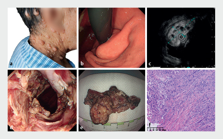 Fig. 1
- —Natural Science Foundation of Henan Province10.13039/501100006407
Peer Reviews
No public reviews on file for this paper yet. If you reviewed it on a platform where reviews are public (OpenReview, ICLR, NeurIPS, ICML), you can paste yours below so the community can read it here.
Videos
No videos yet. Explain this paper in a talk, walkthrough, or lecture? Add one.
Taxonomy
TopicsNeurofibromatosis and Schwannoma Cases · Gastrointestinal Tumor Research and Treatment · Sarcoma Diagnosis and Treatment
A 72-year-old woman was admitted to our hospital with a 20-year history of intermittent subxiphoid pain, which intensified in the past 2 months. Physical examination revealed dermal manifestations suggestive of neurofibromatosis ( Fig. 1 a). Enhanced computed tomography identified thickening of the distal esophageal wall. During upper endoscopy, a mass was observed stretching from the lower esophagus to the gastric cardia, leading to partial esophageal obstruction ( Fig. 1 b). Endoscopic ultrasound depicted a hypoechoic submucosal mass, distinguished by several anechoic lesions extending into the esophageal lumen ( Fig. 1 c). With informed consent from the patient, the mass was successfully excised using endoscopic submucosal dissection ( Fig. 1 d, Video 1 ). The extracted specimen measured 3.0 × 5.0 × 5.0 cm ( Fig. 1 e). Histopathological evaluation confirmed neurofibromatosis, identified by neurofibromatous cells encircling the specimen ( Fig. 1 f). The patient experienced no symptoms and showed no signs of recurrence or residual mass on follow-up endoscopic evaluations over 1 year.
a Cutaneous manifestations of neurofibromatosis observed during physical examination. b Upper endoscopy showing a mass originating from the lower esophagus to the gastric cardia. c Hypoechoic, submucosally-originating mass with multiple anechoic projections into the esophageal cavity, as revealed by endoscopic ultrasound. d Complete removal of the mass via endoscopic submucosal dissection. e The resected specimen. f Histological analysis confirming the presence of neurofibromatosis.
Endoscopic submucosal dissection for a rare cause of esophageal mass: Von Recklinghausen's neurofibromatosis.Video 1
Von Recklinghausen's neurofibromatosis, an autosomal dominant condition, is generally characterized by dermal neurofibromas and distinctive café-au-lait spots. Though predominantly affecting the jejunum, stomach, ileum, and colorectum, esophagus involvement is scarce 1 2 . To the best of our knowledge, this is the inaugural report of an esophageal neurofibromatosis case managed with the ESD technique, demonstrating its safety and efficacy. This case underscores the need for endoscopists to be vigilant for signs of gastrointestinal neurofibroma, particularly in patients with dermal nodules, and suggests that endoscopic approaches like endoscopic submucosal dissection can provide an accurate diagnosis and effective treatment.
Endoscopy_UCTN_Code_TTT_1AO_2AG_3AD
The reference list from the paper itself. Each links out to its DOI / PubMed record.
- 1Tanaka M Kataoka H Joh T Neurofibroma of the esophagus complicating Von Recklinghausenʼs neurofibromatosis Am J Gastroenterol 20131081935193610.1038/ajg.2013.29724300874 · doi ↗ · pubmed ↗
- 2Samat SH Onyemkpa C Torabi M Understanding esophageal neurofibroma: A case series and systematic review Int J Surg Case Rep 20207645045710.1016/j.ijscr.2020.10.03133207410 PMC 7586048 · doi ↗ · pubmed ↗