Beware the glowing fat: pericarditis pitfall
André Vaz

Abstract
Genes, proteins, chemicals, diseases, species, mutations and cell lines named across the full text — each resolved to its canonical identifier and authoritative record.
Click any figure to enlarge with its caption.
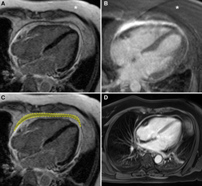 Figure 1
 Figure 2
Peer Reviews
No public reviews on file for this paper yet. If you reviewed it on a platform where reviews are public (OpenReview, ICLR, NeurIPS, ICML), you can paste yours below so the community can read it here.
Videos
No videos yet. Explain this paper in a talk, walkthrough, or lecture? Add one.
Taxonomy
TopicsPericarditis and Cardiac Tamponade · Myasthenia Gravis and Thymoma · Infective Endocarditis Diagnosis and Management
Pericardial inflammation in acute and recurrent pericarditis induces increased vascularity, neutrophil infiltration, and fibrin deposition that, in turn, can be detected by magnetic resonance imaging (MRI) using late gadolinium enhancement (LGE) sequences.^1^ A recent study presented by Cremer et al. elucidated the role of cardiac MRI in assessing recurrent pericarditis and its potential impact on rilonacept treatment duration.^2^ The authors aptly demonstrated the significance of pericardial LGE in predicting the frequency and timing of pericarditis recurrence, emphasizing the importance of tailored therapeutic strategies based on MRI findings.
Phase-sensitive inversion recovery sequences with an inversion time selected for optimal myocardial nulling were used to detect and grade pericardial LGE in the RHAPSODY trial.^2^ However, I would like to point out that while LGE sequences are sensitive to inflammatory and fibrotic changes, their original design without fat saturation may inadvertently highlight epicardial fat, mimicking enhancement (Figure 1A).^3^ Fat usually exhibits a hyperintense signal during the inversion time optimized to null the myocardial signal. LGE sequences with fat saturation are proving particularly useful in this context (Figure 1B), enabling more accurate characterization of true pericardial enhancement (Figure 1C). In situations lacking fat-saturated LGE sequences, Dixon-type sequences (Figure 1D), though not gated, offer an alternative by subtracting fat signal to detect pericardial enhancement. MRI assessment of the pericardium is becoming increasingly common, and a comprehensive understanding of tissue behaviour across different sequences is critical for accurate interpretation of imaging findings.
Lead author biography
André Vaz is a cardiovascular radiologist based in São Paulo, Brazil. He completed his Fellowship in Cardiovascular CT and MRI at the Instituto do Coração do Hospital de Clínicas da Faculdade de Medicina da Universidade de São Paulo, under the supervision of Carlos Eduardo Rochitte. Prior to this, he earned a Master’s Degree in Internal Medicine from the Universidade Federal do Paraná and completed fellowships in Pediatric Radiology at Hospital Pequeno Príncipe. He obtained his Doctor of Medicine degree from the Universidade Federal de Santa Catarina. He is currently involved in research into congenital heart disease, 3D printing, and genetic cardiomyopathies.
The reference list from the paper itself. Each links out to its DOI / PubMed record.
- 1Antonopoulos AS, Vrettos A, Androulakis E, Kamperou C, Vlachopoulos C, Tsioufis K et al Cardiac magnetic resonance imaging of pericardial diseases: a comprehensive guide. Eur Heart J Cardiovasc Imaging 2023;24:983–98.37207354 10.1093/ehjci/jead 092 · doi ↗ · pubmed ↗
- 2Wang TKM, Ayoub C, Chetrit M, Kwon DH, Jellis CL, Cremer PC et al Cardiac magnetic resonance imaging techniques and applications for pericardial diseases. Circ Cardiovasc Imaging 2022;15:e 014283.35861978 10.1161/CIRCIMAGING.122.014283 · doi ↗ · pubmed ↗
- 3Cremer PC, Lin D, Luis SA, Petersen J, Abbate A, Jellis CL et al Pericardial late gadolinium enhancement and time to recurrence: a substudy from RHAPSODY, a phase 3 clinical trial of rilonacept in recurrent pericarditis. Eur Heart J Imaging Methods Pract 2023;1:qyad 003.