Mathematical-Physics Analyses of the Nozzle Shaping at the Aperture Gas Outlet into Free Space under ESEM Pressure Conditions
Pavla Šabacká, Jiří Maxa, Jana Švecová, Jaroslav Talár, Tomáš Binar, Robert Bayer, Petr Bača, Petra Dostalová, Jiří Švarc

TL;DR
This paper studies how nozzle shape affects gas flow and electron beam dispersion in an ESEM under low-pressure conditions.
Contribution
The novelty lies in combining experimental and mathematical-physics approaches to analyze nozzle shaping effects on electron beam dispersion in ESEM.
Findings
Nozzle shape significantly influences supersonic flow and shock wave formation beyond the aperture.
Shock waves impact electron scattering, thereby affecting image quality in ESEM.
Low-pressure conditions are critical in determining the behavior of the electron beam dispersion.
Abstract
The paper presents a methodology that combines experimental measurements and mathematical-physics analyses to investigate the flow behavior in a nozzle-equipped aperture associated with the solution of its impact on electron beam dispersion in an environmental scanning electron microscope (ESEM). The shape of the nozzle significantly influences the character of the supersonic flow beyond the aperture, especially the shape and type of shock waves, which are highly dense compared to the surrounding gas. These significantly affect the electron scattering, which influences the resulting image. This paper analyzes the effect of aperture and nozzle shaping under specific low-pressure conditions and its impact on the electron dispersion of the primary electron beam.
Genes, proteins, chemicals, diseases, species, mutations and cell lines named across the full text — each resolved to its canonical identifier and authoritative record.
Click any figure to enlarge with its caption.
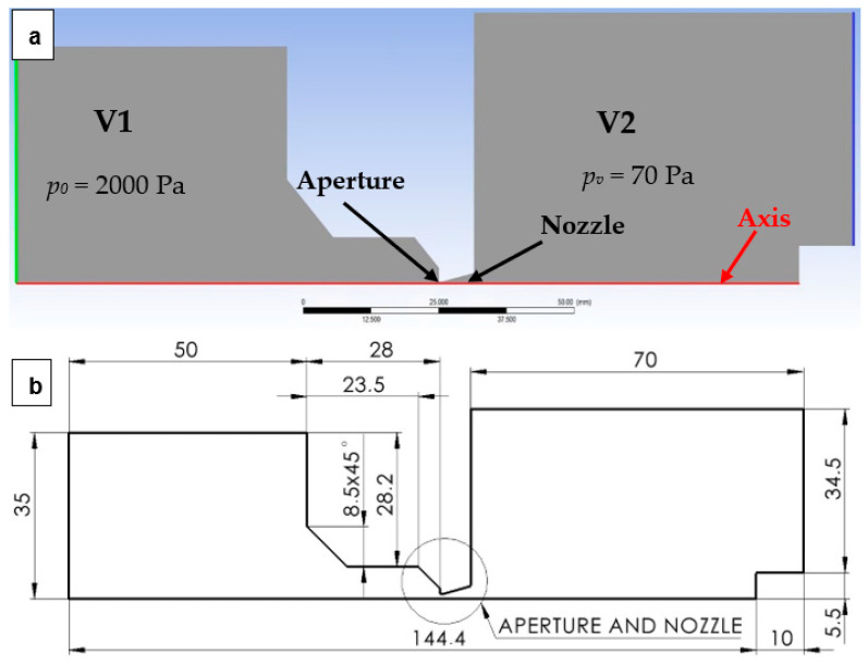 Figure 1
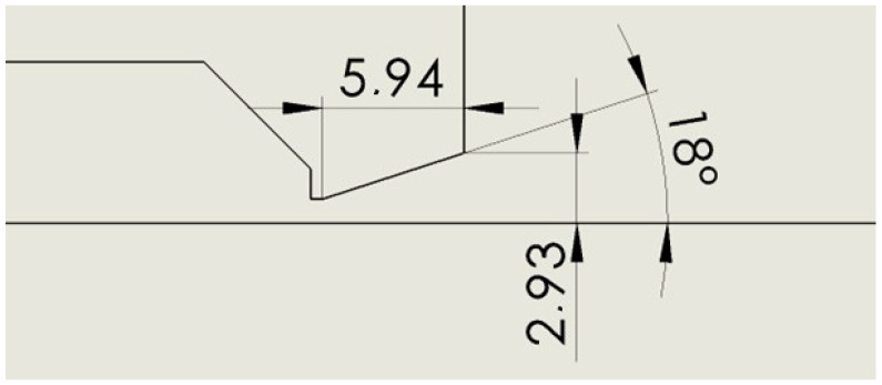 Figure 2
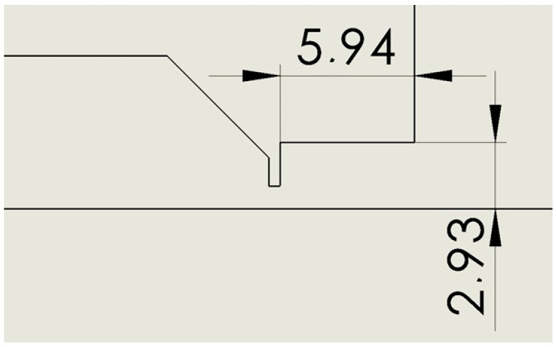 Figure 3
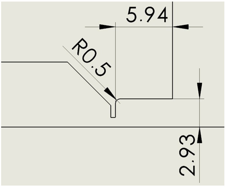 Figure 4
 Figure 5
 Figure 6
 Figure 7
 Figure 8
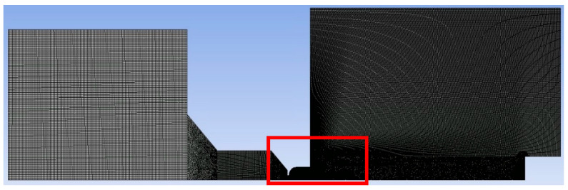 Figure 9
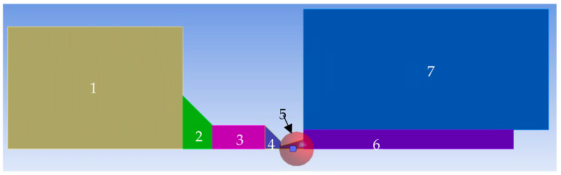 Figure 10
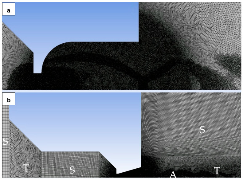 Figure 11
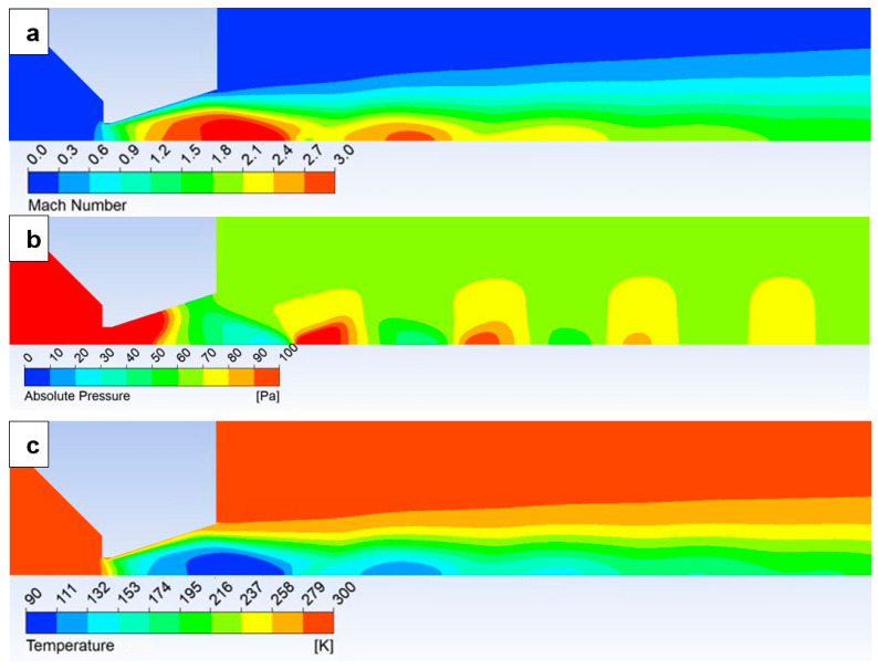 Figure 12
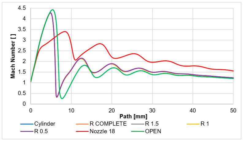 Figure 13
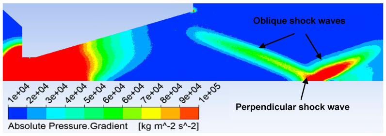 Figure 14
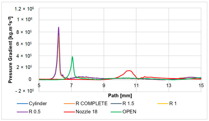 Figure 15
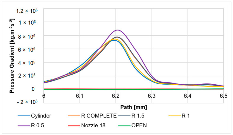 Figure 16
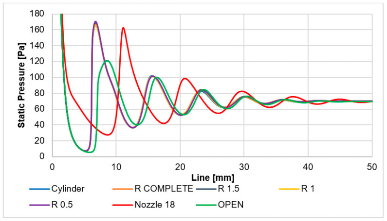 Figure 17
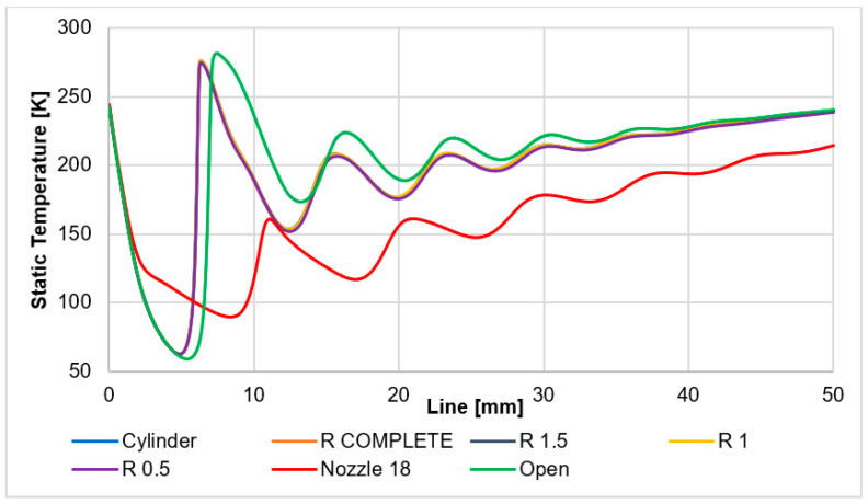 Figure 18
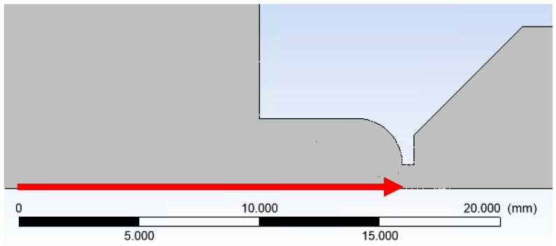 Figure 19
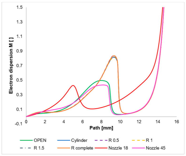 Figure 20
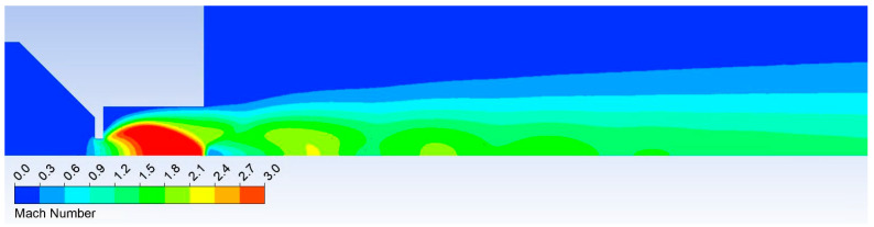 Figure 21
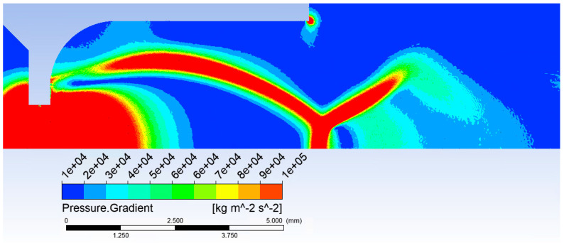 Figure 22
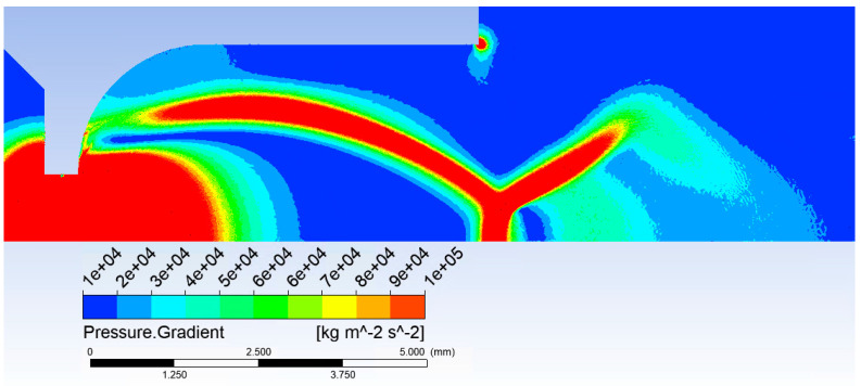 Figure 23
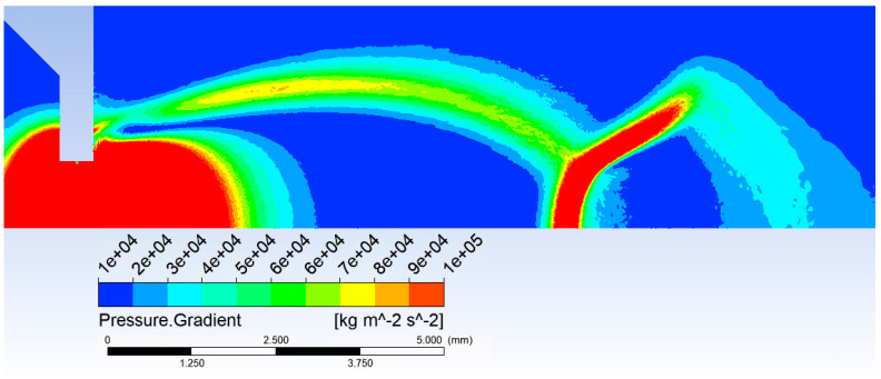 Figure 24
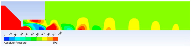 Figure 25
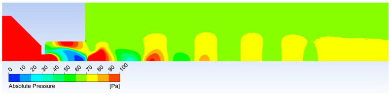 Figure 26
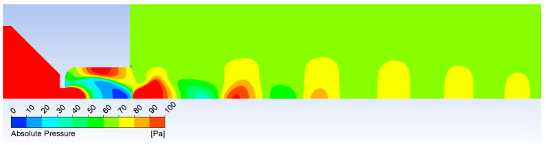 Figure 27
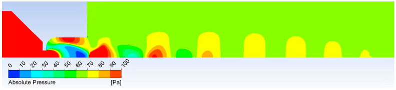 Figure 28
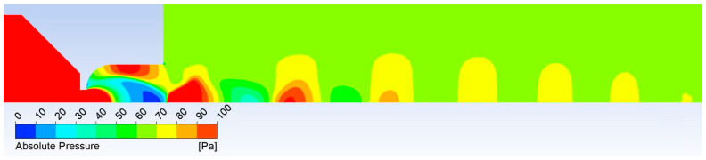 Figure 29
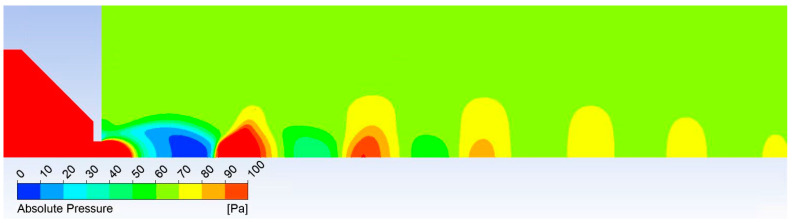 Figure 30
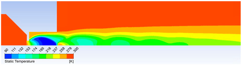 Figure 31
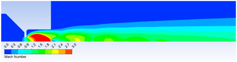 Figure 32
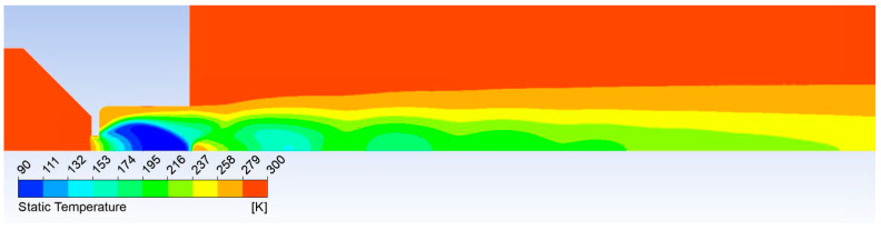 Figure 33
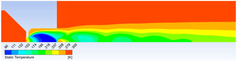 Figure 34
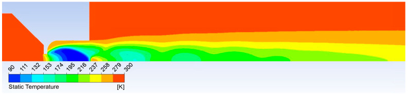 Figure 35
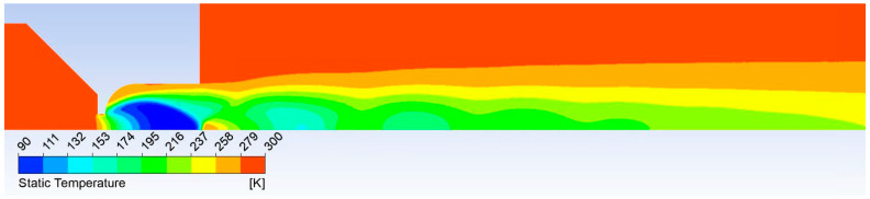 Figure 36
 Figure 37
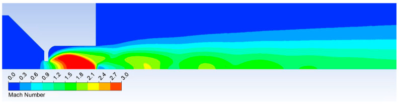 Figure 38
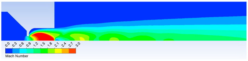 Figure 39
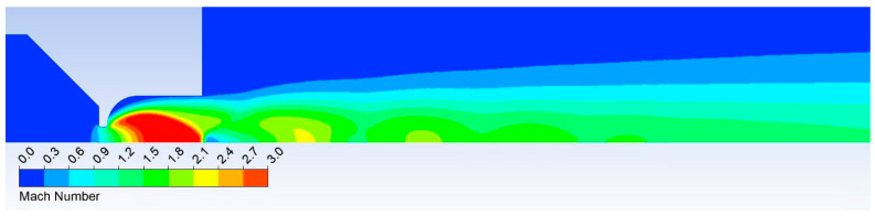 Figure 40
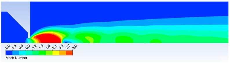 Figure 41
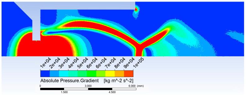 Figure 42
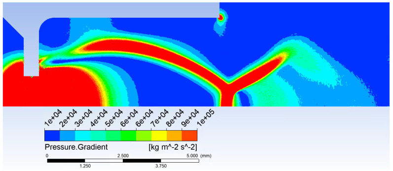 Figure 43
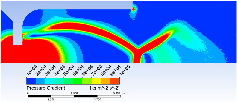 Figure 44
Peer Reviews
No public reviews on file for this paper yet. If you reviewed it on a platform where reviews are public (OpenReview, ICLR, NeurIPS, ICML), you can paste yours below so the community can read it here.
Videos
No videos yet. Explain this paper in a talk, walkthrough, or lecture? Add one.
Taxonomy
TopicsElectron and X-Ray Spectroscopy Techniques · Advancements in Photolithography Techniques · Advanced Electron Microscopy Techniques and Applications