Metabolic Profiling for Unveiling Mechanisms of Kushenol F against Imiquimod-Induced Psoriasis with UHPLC/MS Analysis
Xingxin Yang, Jiaoli Cheng, Xunqing Yin, Ting Ao, Xudong He, Yaqin Yang, Yuping Lin, Zhen Chen

TL;DR
This study shows that Kushenol F reduces psoriasis-like symptoms in mice by modulating inflammation and metabolic pathways.
Contribution
The study identifies 161 metabolites and their pathways affected by Kushenol F in psoriasis treatment.
Findings
Kushenol F reduced PASI scores, epidermal thickening, and inflammatory cytokines in psoriasis-like skin lesions.
Kushenol F regulated 161 metabolites involved in sphingolipid, linoleic acid, and steroid hormone metabolism.
The compound increased IL-10 levels and inhibited pro-inflammatory cytokines like IL-1β, IL-6, and TNF-α.
Abstract
Psoriasis is a common chronic immune-mediated inflammatory skin disorder. Sophora flavescens Alt. (S. flavescens) has been widely acknowledged in the prevention and treatment of psoriasis. Kushenol F (KSCF) is a natural isopentenyl flavonoid extracted from the root of S. flavescens. We aimed to investigate the effect and mechanism of KSCF on imiquimod (IMQ)-induced psoriasis-like skin lesions in mice. A mouse model of psoriasis was induced with 5% IMQ for 5 days, and the mice were given KSCF dermally for 5 days. Changes in skin morphology, the psoriasis area, the severity index (PASI), and inflammatory factors of psoriasis-like skin lesions were evaluated. Metabolites in the psoriasis-like skin lesions were analyzed with ultra-high-performance liquid chromatography/mass spectrometry followed by a multivariate statistical analysis to identify the differential metabolites and metabolic…
Genes, proteins, chemicals, diseases, species, mutations and cell lines named across the full text — each resolved to its canonical identifier and authoritative record.
Click any figure to enlarge with its caption.
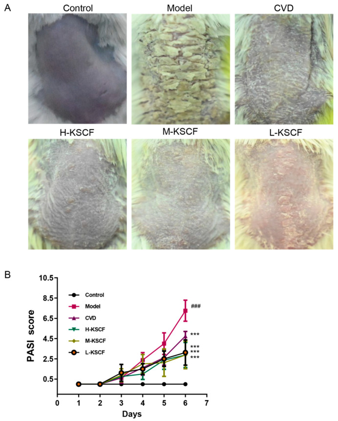 Figure 1
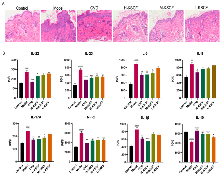 Figure 2
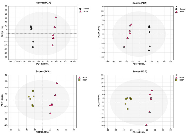 Figure 3
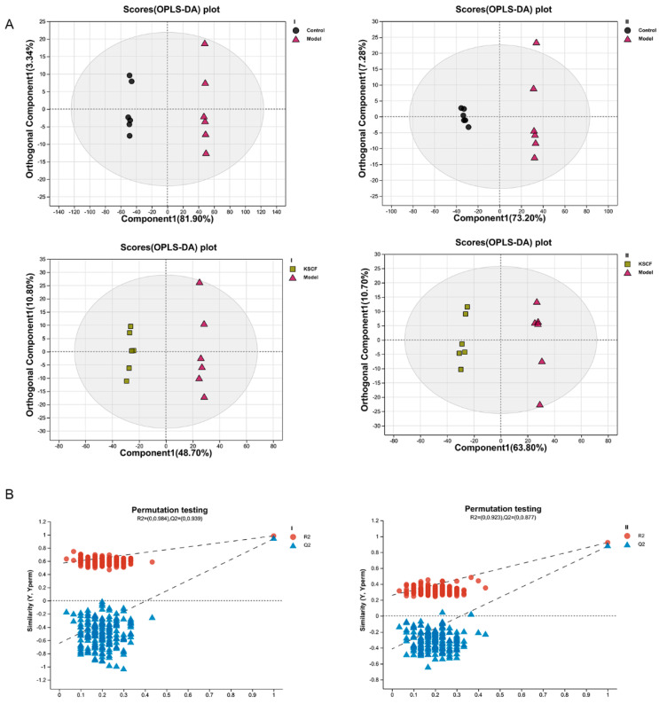 Figure 4
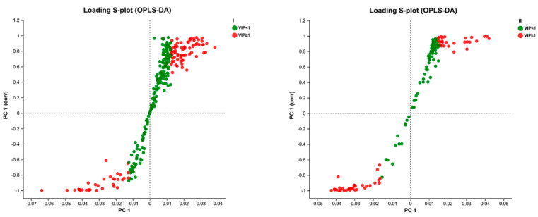 Figure 5
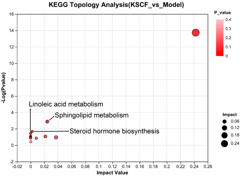 Figure 6
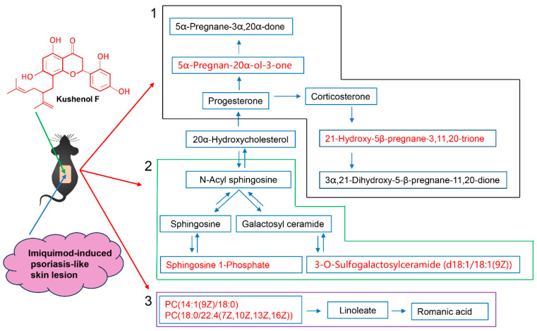 Figure 7
- —National Natural Science Foundation of China
- —Application and Basis Research Project of Yunnan China
Peer Reviews
No public reviews on file for this paper yet. If you reviewed it on a platform where reviews are public (OpenReview, ICLR, NeurIPS, ICML), you can paste yours below so the community can read it here.
Videos
No videos yet. Explain this paper in a talk, walkthrough, or lecture? Add one.
Taxonomy
TopicsPsoriasis: Treatment and Pathogenesis · Phytochemistry Medicinal Plant Applications · Synthesis and biological activity
1. Introduction
Psoriasis is a chronic and systemic immune-mediated disease. The underlying pathomechanisms involve an interplay between the innate and adaptive immune systems [1,2]. Recently, psoriasis presents many challenges, including high prevalence, chronicity, and associated comorbidity [3]. As a representative inflammatory skin disease occupied by large surface involvement, the occurrence and development of psoriasis is reported to be related to the disorder of many inflammation markers, such as IL-17A, IL-23, and TNF-α. Biologics targeting IL-17A, IL-23, and TNF-α have been developed and approved for the treatment of psoriasis [4]. The progression of psoriasis has also been confirmed to be related to immunity [5,6,7].
Common treatments for psoriasis include topical agents, systemic therapy, and ultraviolet light therapy. Topical agents mainly include calcipotriol (CVD), corticosteroids, and salicylic acid. Methotrexate, cyclosporine, and acitretin are commonly used as systemic therapy for psoriasis [8,9]. Although the therapeutic effects of these drugs have been widely recognized, adverse reactions still largely limit their clinical utilization [10]. Hence, complementary and alternative medicine may offer options for some patients.
Sophora flavescens Alt. (S. flavescens) is widely used in the clinic, can clear dampness-heat, kill insects, and dispel wind evil, which originates from the teachings of traditional Chinese medicine [11]. S. flavescens has been clinically used in numerous countries to treat skin diseases [12,13]. Kushenol F (KSCF) is a natural isopentenyl flavonoid extracted from the root of S. flavescens. It has been reported to have anti-inflammatory and anti-bacterial properties [14,15,16]. A previous study demonstrated that KSCF oral treatment could be an important therapeutic for treating atopic dermatitis. Although various studies have reported the biological effects of KSCF, the effect and mechanism of KSCF on psoriasis are unclear.
Metabolomics can reveal the specific changes in physiological and biochemical states related to phenotypes in biological systems. The application of skin metabonomics is suitable for revealing the potential functional changes in a variety of metabolic pathways and signal networks in psoriasis-like organisms, which has become a significant approach to evaluate the efficacy and mechanism of drugs [17]. The present study investigated the effects of KSCF on imiquimod (IMQ)-induced psoriasis-like skin lesions in mice using conventional pharmacology and metabolomics. The potential biomarkers and complex mechanisms of KSCF involved in psoriasis treatment are also discussed. The results demonstrated that KSCF inhibited the inflammatory response to prevent IMQ-induced psoriasis-like skin lesions in mice by call-backing the levels of 161 endogenous metabolites and affecting their related metabolic pathways. Thus, KSCF has the potential to be developed as a topical drug for treating psoriasis symptoms.
2. Results and Discussion
2.1. KSCF Mitigated the Psoriatic Dermatitis Induced by IMQ
Psoriasis is a skin disease with characteristics of well-demarcated, erythematous, raised lesions with silvery-white dry scales. As depicted in Figure 1, after 5 days of IMQ treatment, erythema and silvery-white dry scales were observed on the dorsal skin, and the PASI was significantly increased (p < 0.001). However, after treatment with CVD and KSCF, the thickness and size of the scales on the dorsal skin were decreased. Erythema and PASI were also significantly decreased (p < 0.001). These results indicate that KSCF effectively relieved IMQ-induced psoriasis symptoms.
2.2. Effect of KSCF on Inflammatory Reactions in Psoriasis-like Mice
Psoriasis is a chronic inflammatory skin disease characterized by lymphocyte and neutrophil infiltration and the excessive keratinization and proliferation of keratinocytes [18,19]. The agents capable of attenuating keratinocyte hyperproliferation and excessive inflammatory responses are considered to be potentially useful for psoriasis treatment [20,21]. IMQ-induced psoriasis is usually accompanied by cellular inflammatory factor disorders in the dorsal skin. As shown in Figure 2A, IMQ induced massive inflammatory cell infiltration in the dorsal skin, which was reduced by CVD and KSCF treatment. As shown in Figure 2B, after 5 days of IMQ treatment, the levels of IL-1β, IL-6, IL-8, IL-17A, IL-22, IL-23, and TNF-α significantly increased in the dorsal skin (p < 0.05), whereas IL-10 levels significantly decreased (p < 0.0001). After treatment with CVD and KSCF, the levels of IL-1β, IL-6, IL-8, IL-17A, IL-22, IL-23, and TNF-α significantly decreased (p < 0.05) and IL-10 levels significantly increased (p < 0.0001). These results indicated that KSCF might stimulate the secretion of anti-inflammatory cytokines while inhibiting the release of pro-inflammatory cytokines, thereby alleviating IMQ-induced psoriasis.
2.3. Effect of KSCF on Metabolites in the Injured Skin of Psoriasis-like Mice
The data of skin samples were collected with ultra-high-performance liquid chromatography/mass spectrometry (UHPLC/MS), and the metabolic profiles of each group were obtained. As depicted in Supplementary Figure S1, the peak number and intensity significantly varied in the total ion current profiles of the control, model, CVD, and KSCF groups, indicating significant differences among the metabolic state of each group and endogenous metabolites. Thus, the endogenous metabolites of skin changed significantly after IMQ induction and administration. In this study, PCA and OPLS-DA were used to analyze metabolome differences in the dorsal skin between the control, model, and KSCF groups using an unsupervised statistical method. The metabolic state of the control and model groups was different (Figure 3 and Figure 4A). The KSCF group was far from the model group. The results demonstrated that KSCF effectively reversed the pathological changes triggered by IMQ treatment in psoriasis-like mice.
Two hundred iteration permutation tests were performed on OPLS-DA in the positive and negative ion modes to further illustrate the reliability of the OPLS-DA model. The Q^2^ values were less than 0.05, further confirming the accuracy of the multivariate statistical analysis (Figure 4B).
2.4. Identification of Differential Metabolites
Changes in the metabolites can reflect the physiological and pathological states of the human body and might serve as an objective index to determine the efficacy and mechanism of drugs during disease intervention [22]. The complete metabolic information was collected, and the mechanism of KSCF against IMQ-induced psoriasis, which involved the regulation of endogenous metabolites, was revealed using the UHPLC/MS technique. S-plot diagrams based on OPLS-DA analysis were plotted to obtain information on differential metabolites between the model group and the KSCF group. Substances with a VIP of >1 and p-value of <0.05 were selected as biomarkers (Figure 5). A total of 161 potential biomarkers were detected in the dorsal skin, of which 102 metabolites were up-regulated, and 59 metabolites were down-regulated (Table 1; their chemical structures are shown in Supplementary Figure S2). These results suggested that KSCF might regulate these differential metabolites to relieve IMQ-induced psoriasis.
2.5. Metabolic Pathway Analysis
The differential metabolites were subjected to a pathway enrichment analysis to explore the potential metabolic pathways of KSCF exerting medicinal effects. Figure 6 shows the path influence diagrams of the metabolic pathway analysis. Pathways with a p-value of <0.05 were considered KSCF-involved pathways. The difference between the model and KSCF groups was clearly reflected in three pathways (Table 2), which are mainly related to lipid metabolism and steroidogenic activity.
Sphingolipids have structural and biological functions in the human epidermis, are importantly involved in the maintenance of the skin barrier, and regulate cellular processes, such as the proliferation, differentiation, and apoptosis of keratinocytes [23,24]. In this study, 3-O-sulfogalactosylceramide (d18:1/18:1(9Z)) and sphingosine 1-phosphate were identified as metabolites with significant differences after modeling and drug administration, suggesting that these key metabolites could be targets for KSCF against IMQ-induced psoriasis. Therefore, it was inferred that KSCF improved IMQ-induced psoriasis by regulating sphingolipid metabolism pathways.
Abnormal linoleic acid metabolism has been shown to be a key pathway in psoriasis. The combination of a moisturizer containing a linoleic acid–ceramide complex and glucocorticoids was shown to significantly improve the therapeutic efficacy of psoriasis and delay recurrence [25]. Our results confirmed that the levels of PC (14:1(9Z)/18:0) and PC (18:0/22:4(7Z,10Z,13Z,16Z)) were close to the normal group after KSCF administration. These results suggested that KSCF improved IMQ-induced psoriasis by regulating the linoleic acid metabolism pathways.
The skin has an endogenous steroidogenic capacity, and modulating local steroidogenic activity may be a new approach to treating inflammatory skin diseases [26,27]. In this study, 5α-Pregnan-20α-ol-3-one and 21-Hydroxy-5b-pregnane-3,11,20-trione were identified as metabolites with significant differences after modeling and drug administration. After KSCF administration, steroid hormone biosynthesis was similar to the normal group, suggesting that KSCF treatment affected steroid hormone biosynthesis to relieve IMQ-induced psoriasis.
Our results indicated that KSCF inhibited the inflammatory response to prevent IMQ-induced psoriasis-like skin lesions in mice by call-backing the levels of endogenous metabolites and affecting their related metabolic pathways (Figure 7). It is very likely that the flavonoid extract from S. flavescens also shows the therapeutic effects on psoriasis because it contains many active compounds, such as KSCF, which is worthy of further study. The deep molecular mechanism of KSCF regulating endogenous metabolites in three key pathways is also worthy to be further explored by detecting the key proteins in the pathways. It is reported that another flavonoid (kurarinone) and alkaloids (matrine and oxymarine) isolated from S. flavescens regulated the inflammatory response to intervene in psoriasis [7,28,29]. However, the differences in the efficacy and mechanism of these compounds in the psoriasis treatment and the relationship between the chemical structures of them and their anti-psoriasis activity are still unclear, which is worthy of further study.
3. Materials and Methods
3.1. Chemicals, Reagents, and Materials
KSCF was provided from Chengdu Pufei De Biotech Co., Ltd. (Chengdu, China). CVD was purchased from A&M Pharmaceuticals (Hong Kong, China). Hematoxylin stain was obtained from Wuhan Google Biotechnology Co., Ltd. (Wuhan, China). Vaseline for medical use was purchased from Shandong Lircon Medical Technology Co., Ltd. (Dezhou, China). IMQ was acquired from Sichuan MED-SHINE Pharmaceutical Co., Ltd. (Chengdu, China). Four percent paraformaldehyde solution was provided from Beijing lanjieke Technology Co., Ltd. (Beijing, China). IL-6, IL-8, IL-1β, IL-10, IL-22, IL-23, IL-17A, and TNF-α ELISA Kits were purchased from Jiangsu Meimian Industrial Co., Ltd. (Yancheng, China). The reference substance of KSCF (purity > 98%) was purchased from Chengdu Pufei De Biotech Co., Ltd. (Chengdu, China). High purity deionized water was purified using a Milli-Q system (Millipore, Bedford, MA, USA). HPLC-grade formic acid, methyl alcohol, and acetonitrile were acquired from Fischer Control Equipment International Co., Ltd. (Hong Kong, China). H&E Staining kits were purchased from Beijing Solarbio Science&Technology Co., Ltd. (Beijing, China). Seventy-five percent ethanol was purchased from Sinopharm Chemical Reagent Co., Ltd. (Shanghai, China). All other reagents used were of analytical reagent grade or higher.
3.2. Animals and Experimental Protocol
All experimental procedures in this study complied with the National Guidelines for the Care and Use of Laboratory Animals and were approved by the Animal Ethics Committee of Yunnan University of Chinese Medicine, with ethics number R062021158.
Healthy male BALB/c mice (20 ± 2 g), aged 6–8 weeks, were obtained from Sipeifu Biotechnology Co., Ltd. (Beijing, China). The mice were kept in the Laboratory of Yunnan University of Traditional Chinese Medicine (Kunming, China), following standard housing conditions. All mice were adaptively fed for one week. The mice were randomized into six groups (n = 6 mice per group), including control group (75% ethanol), model group (75% ethanol), positive group (CVD, 62.5 mg/kg), low-dose KSCF group (L-KSCF, 200 mg/kg), medium-dose KSCF group (M-KSCF, 400 mg/kg), and high-dose KSCF group (H-KSCF, 600 mg/kg). Except for the control group, mice were topically treated with 5% IMQ cream at a daily dose of 62.5 mg for a duration of 5 days on their dorsal region. KSCF and CVD were dissolved in 75% ethanol. Starting from the day of modeling, the corresponding drugs were sprayed continuously for 5 days (0.3 mL/day/mouse) in the administration group. Seventy-five percent ethanol (0.3 mL/day/mouse) was sprayed for the control group and the model group.
3.3. Psoriasis Area and Severity Index (PASI) Assessment
According to clinical PASI score standard, the severity of the skin inflammation was evaluated once daily, including the measurements for skin erythema, scaling, and thickening. Erythema, scaling, and thickening were scored independently on a scale from 0 to 4: 0, none; 1, slight; 2, moderate; 3, marked; 4, very marked. The cumulative score (erythema plus scaling plus thickening) served as a measure of the severity of inflammation (scale 0–12) [30].
3.4. Image Acquisition and Skin Sample Collection
After euthanizing the mice through carbon dioxide asphyxiation, images were captured on the dorsal side of each mouse group. Then, the dorsal skin of the mice was carefully excised using surgical scissors and divided into two sections. One section was immediately immersed in liquid nitrogen for cryopreservation, while the other section was fixed in 4% paraformaldehyde universal tissue fixative.
3.5. Histopathology Analyses
The dorsal skin of mice was fixed in 4% paraformaldehyde universal tissue fixative for 24 h, washed with PBS, dehydrated with gradient ethanol and transparentized with xylene, and embedded in paraffin. After hematoxylin and eosin staining, histological parameters were observed under a light microscope, and images were taken at 200× magnification.
3.6. Measurement of Skin Inflammatory Factors
Approximately 50 mg of injured skin from the dorsal region of each mouse were weighed and placed on ice. The surface blood stains on the skin samples were rinsed with pre-cooled normal saline solution. After air-drying the filter paper, the skin samples were promptly sectioned into pieces and transferred into a covered 2 mL centrifuge tube. The tube was then subjected to centrifugation (Centrifuge 5430R, Eppendorf, Germany) at 1500× g for 15 min, and subsequently, the supernatant of the homogenized tissue was collected for further utilization. The levels of cytokines IL-1β, IL-6, IL-8, IL-10, IL-17A, IL-22, IL-23, and TNF-α in the dorsal skin lesions of all groups of mice were detected according to the kit manufacturer’s instructions. The values were determined by measuring the absorbance value (OD) at 450 nm using a microplate reader (Rayto Life and Analytical Sciences Co., Ltd., Shenzhen, China).
3.7. Metabolomic Analysis
3.7.1. Sample Pre-Treatment
Skin samples: 50 mg skin sample and a 6 mm diameter grinding bead was added to a 2 mL centrifuge tube. An amount of 400 μL of extraction solution (methanol: water = 4:1 (v:v)) was used for metabolite extraction. Samples were ground with Wonbio-96c frozen tissue grinder (Shanghai Wanbo Biotechnology Co., Ltd., Shanghai, China) for 6 min (−10 °C, 50 Hz) followed by low-temperature ultrasonic extraction for 30 min (5 °C, 40 kHz). Then, the sample was allowed to stand for 30 min at −20 °C and centrifuged for 15 min (4 °C, 3500× g). The supernatant was transferred to a clean tube and dried gently with nitrogen. The residues were redissolved in 200 µL of methanol for UHPLC/MS analysis.
Quality control sample: A random injection sequence was employed to detect signal fluctuation. An amount of 2 μL of skin samples were taken and thoroughly mixed. Then, the mixture was centrifuged at 3500× g for 15 min at 4 °C. The supernatant was collected to perform UHPLC/MS analysis.
3.7.2. UHPLC/MS Analysis
UHPLC analyses were performed using Ultimate 3000 Ultra High Performance Liquid Chromatography (Thermo Fisher Scientific, San Jose, CA, USA) equipped with a Thermo Scientific Hypersil GOLD column (50 mm × 2.1 mm, 1.9 μm). The column temperature was maintained at 40 °C to obtain better sample separation. The mobile phases consisted of water containing 0.1% formic acid (A) and acetonitrile (B) at a 0.4 mL/min of flow rate. The gradient program was set as follows: 0–3.5 min, 0% B → 24.5% B; 3.5–5 min, 24.5% B → 65% B; 5–7.4 min, 65% B → 100% B; 7.4–7.6 min, 100% B → 51.5% B; 7.6–10 min, 51.5% B → 0% B.
The MS data were collected using a Thermo Scientific Q-Exactive TM hybrid quadrupole-orbitrap mass spectrometer with a heated electrospray ionization probe (Thermo Fisher Scientific, San Jose, CA, USA). MS conditions were as follows: CUR, 15 psi; Gas1 and Gas2, 50 psi; IS, 5500 V; gas temperature, 500 °C.
3.8. Statistical Analysis
All UHPLC/MS raw files were exported in comma-separated value (CSV) format using the Progenesis QI (Waters Corporation, Milford, CT, USA) software. All data were uploaded to XCMS online for peak alignment, normalization, and retrieval. The data were analyzed with principal component analysis (PCA) and orthogonal partial least squares discriminant analysis (OPLS-DA) using the SIMCA-P 14.1 software (Sweden, Umeå, Umetrics). The potential biomarkers were selected according to the parameters of variable importance in the projection (VIP > 1 and p < 0.05) from OPLS-DA. The structural information of the metabolites was identified using the HMDB databases. Finally, the metabolic pathways were searched using Kyoto Encyclopedia of Genes and Genomes (KEGG) through MetaboAnalyst 5.0 online, and the pathways with an impact value greater than 0.1 were considered KSCF-involved pathways.
All experimental data were presented as means ± S.D. Each experiment was performed in triplicate. The overall significance of the results was examined with one-way ANOVA using GraphPad Prism 5.0 (GraphPad Software, La Jolla, CA, USA). Kolmogorov–Smirnov test was used for the pre-test of the analysis of ANOVA to ensure the rationality of the statistical analysis. Student’s t-test delivered the p-values shown. The differences between the compared groups were considered statistically significant at p < 0.05, p < 0.01, p < 0.001, p < 0.0001.
4. Conclusions
KSCF inhibited the inflammatory response to prevent IMQ-induced psoriasis-like skin lesions in mice by call-backing the levels of 161 endogenous metabolites and affecting the three related metabolic pathways, including sphingolipid and linoleic acid metabolism and steroid hormone biosynthesis. Thus, KSCF has the potential to be developed as a topical drug for treating psoriasis.
The reference list from the paper itself. Each links out to its DOI / PubMed record.
- 1Wu J.J. Kavanaugh A. Lebwohl M.G. Gniadecki R. Merola J.F. Psoriasis and metabolic syndrome: Implications for the management and treatment of psoriasis J. Eur. Acad. Dermatol. Venereol.20223679780610.1111/jdv.1804435238067 PMC 9313585 · doi ↗ · pubmed ↗
- 2Armstrong A.W. Read C. Pathophysiology, Clinical Presentation, and Treatment of Psoriasis: A Review JAMA 2020191945196010.1001/jama.2020.400632427307 · doi ↗ · pubmed ↗
- 3Lee H.J. Kim M. Challenges and future trends in the treatment of psoriasis Int. J. Mol. Sci.2023241331310.3390/ijms 24171331337686119 PMC 10487560 · doi ↗ · pubmed ↗
- 4Tokuyama M. Mabuchi T. New treatment addressing the pathogenesis of psoriasis Int. J. Mol. Sci.202021748810.3390/ijms 2120748833050592 PMC 7589905 · doi ↗ · pubmed ↗
- 5Liu L. Jin R. Hao J. Zeng J. Yin D. Yi Y. Zhu M. Mandal A. Hua Y. Ng C.K. Consumption of the fish oil high-fat diet uncouples obesity and mammary tumor growth through induction of reactive oxygen species in protumor macrophages Cancer Res.2020802564257410.1158/0008-5472.CAN-19-318432213543 PMC 7299802 · doi ↗ · pubmed ↗
- 6Hao J. Jin R. Zeng J. Hua Y. Yorek M.S. Liu L. Mandal A. Li J. Zheng H. Sun Y. Consumption of fish oil high-fat diet induces murine hair loss via epidermal fatty acid binding protein in skin macrophages Cell Rep.20224111180410.1016/j.celrep.2022.11180436516778 PMC 10193786 · doi ↗ · pubmed ↗
- 7Kim B.H. Na K.M. Oh I. Song I.H. Lee Y.S. Shin J. Kim T.Y. Kurarinone regulates immune responses through regulation of the JAK/STAT and TCR-mediated signaling pathways Biochem. Pharmacol.2013851134114410.1016/j.bcp.2013.01.00523333426 · doi ↗ · pubmed ↗
- 8Griffiths C.E.M. Armstrong A.W. Gudjonsson J.E. Barker J.N.W.N. Psoriasis Lancet 20213971301131510.1016/S 0140-6736(20)32549-633812489 · doi ↗ · pubmed ↗