Correction: Inhibition of GSK3β activity alleviates acute liver failure via suppressing multiple programmed cell death
Danmei Zhang, Chunxia Shi, Qingqi Zhang, Yukun Wang, Jin Guo, Zuojiong Gong

Abstract
Genes, proteins, chemicals, diseases, species, mutations and cell lines named across the full text — each resolved to its canonical identifier and authoritative record.
Click any figure to enlarge with its caption.
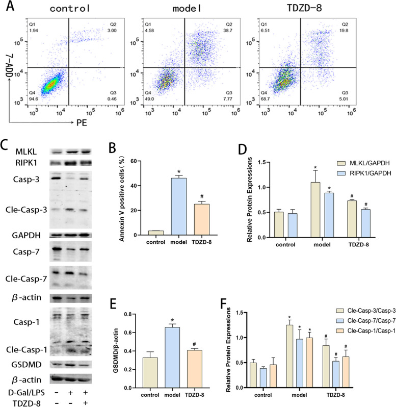 Figure 1
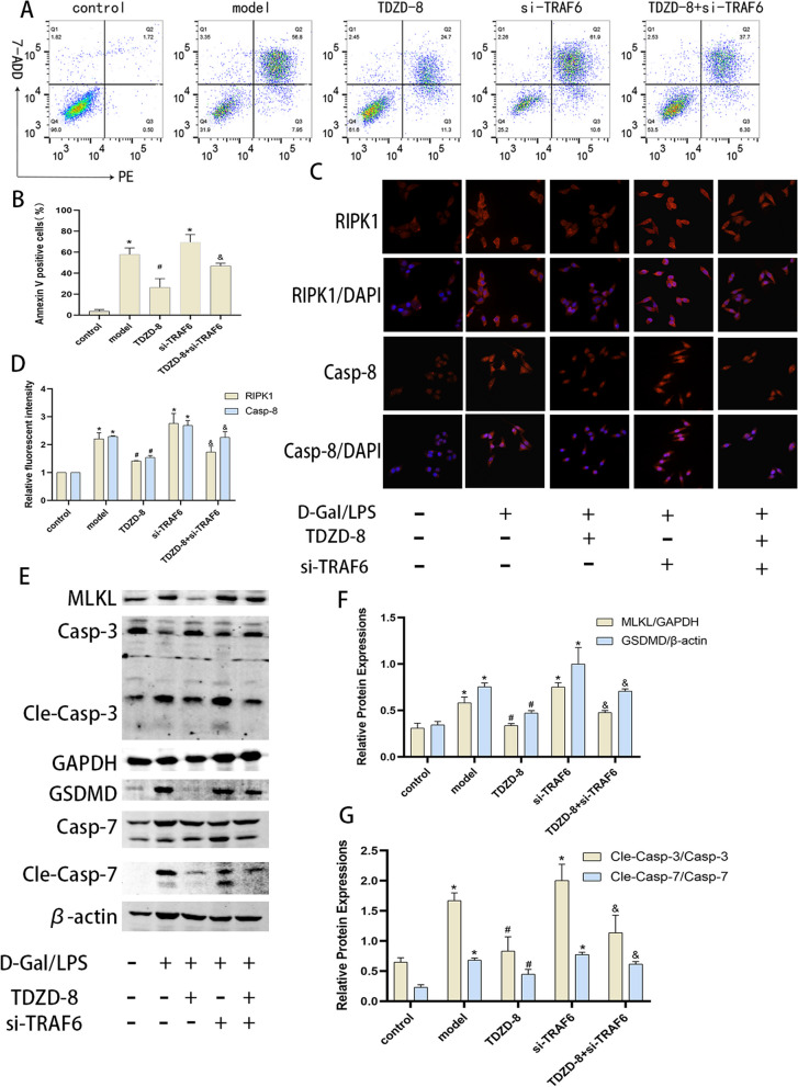 Figure 2
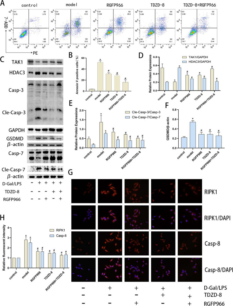 Figure 3
Peer Reviews
No public reviews on file for this paper yet. If you reviewed it on a platform where reviews are public (OpenReview, ICLR, NeurIPS, ICML), you can paste yours below so the community can read it here.
Videos
No videos yet. Explain this paper in a talk, walkthrough, or lecture? Add one.
Taxonomy
TopicsCancer-related gene regulation · Ubiquitin and proteasome pathways · RNA Research and Splicing
Correction: J Inflam 20, 24 (2023)
https://doi.org/10.1186/s12950-023-00350-1
After publication of this article, it was reported that in Figs. 3, 5 and 7, some errors occurred; the figures should have appeared as shown below.
The first is a set of cellular immunofluorescence images of CAS8 molecules in Fig. 5C. The first two merge images were inverted due to negligence. Secondly, regarding the statistical analysis of the apoptosis flow pattern in Figs. 3, 5 and 7, it was not the intention to analyze the early apoptotic cell population as an apoptosis indicator to support the conclusion. However, after carefully discussing and reviewing two instructions from BD and Abcam, and based on previous studies [1, 2], it would be more rigorous to change the vertical coordinate of the graph to Annexin V positive cells (%) instead of apoptosis rate in Figs. 3, 5 and 7, which would only illustrate the results of the experiments.Fig. 3TDZD-8 alleviates the level of death in D-Gal/LPS stimulated cells. A and (B) the Percentage of apoptotic cells detected by flow cytometry. C-F Expression of MLKL, RIPK1, GSDMD, cleaved caspase-7, cleaved caspase-3 and cleaved caspase-1 protein in each group of cells and their quantitative analysis. *P < 0.05 compare with control group, #P < 0.05 compare with model groupFig. 5Inhibition of TRAF6 was able to partially reverse the hepatocyte-protective effect of TDZD-8 in vitro. A and (B) Flow cytometry detection of apoptosis levels in each group and its statistical analysis. C and (D) Immunofluorescence detection of RIPK1 and caspase-8 localization and expression levels in vitro and their quantitative analysis (magnification × 400). E–G Protein blotting and immunofluorescence to detect MLKL, GSDMD, cleaved caspase-7, cleaved caspase-3 protein levels in each group of cells and their quantitative analysis. *P < 0.05 compare with control group, #P < 0.05 compare with model group, &P < 0.05 compare with TDZD-8 groupFig. 7Inhibition of HDAC3 levels modulates TAK1 levels and attenuates the level of death in acute hepatocyte injury. A and (B) the Percentage of apoptotic cells in each group by flow cytometry. C-F Protein blotting of TAK1, HDAC3, GSDMD, cleaved caspase-7 and cleaved caspase-3 protein expression in vitro and quantitative analysis. G and (H) Immunofluorescence detection of RIPK1 and caspase-8 localization and expression in vitro (magnification × 400). *P < 0.05 compare with control group, #P < 0.05 compare with model group
The original article has been updated by the authors.
The reference list from the paper itself. Each links out to its DOI / PubMed record.
- 1Meza-Sosa KFSPARCLE, a p 53-induced lnc RNA, controls apoptosis after genotoxic stress by promoting PARP-1 cleavage Mol Cell 2022824785802.e 1010.1016/j.molcel.2022.01.00135104452 PMC 10392910 · doi ↗ · pubmed ↗
- 2Gourisankar S Rewiring cancer drivers to activate apoptosis Nature 2023620797341742510.1038/s 41586-023-06348-237495688 PMC 10749586 · doi ↗ · pubmed ↗