A Divergent Sequela of Scleral Buckle Removal
Leroy Tan, Fiona Chew Lee Min, Zabri B Kamarudin, Safinaz Mohd Khialdin

TL;DR
A patient developed strabismus after scleral buckle removal and was successfully treated with strabismus surgery.
Contribution
Reports a unique case of strabismus following scleral buckle removal and its successful surgical management.
Findings
Strabismus occurred one month after scleral buckle removal due to anterior migration and exposure.
Strabismus surgery successfully relieved diplopia in the patient's primary gaze.
Abstract
Scleral buckle (SB) removal is done for a variety of reasons following treatment of rhegmatogenous retinal detachments (RRD), such as buckle exposure, migration, and infection. The most worrying complication of SB removal is retinal redetachment. We report a unique case of a patient developing strabismus about one month after scleral buckle removal for anterior migration and exposure of the scleral buckle. We also share a successful strabismus surgery which had a main aim of relieving diplopia in the patient's primary gaze.
Genes, proteins, chemicals, diseases, species, mutations and cell lines named across the full text — each resolved to its canonical identifier and authoritative record.
Click any figure to enlarge with its caption.
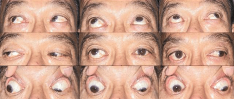 Figure 1
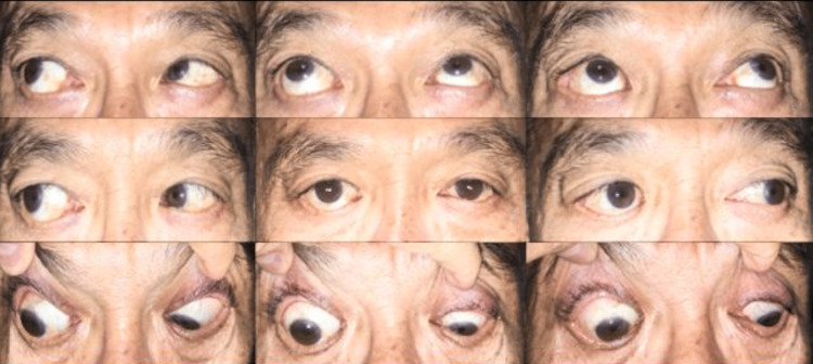 Figure 2
Peer Reviews
No public reviews on file for this paper yet. If you reviewed it on a platform where reviews are public (OpenReview, ICLR, NeurIPS, ICML), you can paste yours below so the community can read it here.
Videos
No videos yet. Explain this paper in a talk, walkthrough, or lecture? Add one.
Taxonomy
TopicsRetinal and Macular Surgery · Intraocular Surgery and Lenses · Glaucoma and retinal disorders
Introduction
Scleral buckle (SB) removal is done for a variety of reasons following treatment of rhegmatogenous retinal detachments (RRD), such as buckle exposure, migration, and infection [1,2]. Strabismus itself can be caused by an in-situ scleral buckle [1]. The most worrying complication of SB removal is retinal redetachment [1]. This case report describes an uncommon development of strabismus in an adult patient as a complication of SB removal.
This case was previously presented as a poster at the 7th USM Ophthalmology Symposium in conjunction with the 9th Conjoint Ophthalmology Scientific Conference, on 13th September 2019.
Case presentation
A 50-year-old gentleman, with no medical illnesses had an encircling SB removal done for RRD in the right eye (RE) 5 years prior and postoperatively enjoyed good vision with the treated eye. He had presented acutely with a complaint of red eye, eye discharge, and pain to the vitreoretinal clinic. He reported no diplopia. At that time, the SB was found to be anteriorly migrated throughout and there was an exposed area at the supero-temporal aspect of the RE. He was admitted for intravenous antibiotics and SB removal was done.
At the one-month follow-up visit after SB removal, he complained of a progressively worsening double vision. On examination, visual acuity was 6/24 and 6/9 in his right and left eye, respectively, with no measurable stereopsis. There was no pain on eye movement. He had an alternating exotropia of 40-prism diopters (PD) and hypertropia of 30-PD. There was also limited depression and adduction of the RE. There were no signs of re-detachment. Figure 1 below shows the 9-gaze view at the one-month follow-up visit after SB removal.
Alternating exotropia with limited depression and adduction of the right eye one month after scleral buckle removal surgery for buckle anterior migration and exposure
Magnetic resonance imaging of the orbits revealed intact extraocular muscles (EOM). His thyroid function tests were normal. Eight months after the initial SB removal, the patient underwent bilateral lateral rectus recession of 7.5mm and RE superior rectus recession of 7.0mm. Intraoperatively, a forced duction test revealed limited adduction and depression. The lateral and superior recti were adherent to the globe with massive fibrosis.
One week after squint surgery, the patient's diplopia resolved, and at the one-year follow-up, he was orthophoric with no EOM restriction and no signs of retinal redetachment. This patient is to be seen at least yearly to monitor for re-detachment. Figure 2 below shows the 9-gaze view at the one-year follow-up visit post-squint surgery.
The patient was orthophoric in primary position at the one-year follow-up visit after squint surgery
Discussion
Strabismus development after buckle surgery for retinal detachment repair is fairly common [2]. Removal of SB is sometimes done to improve strabismus following buckle surgery. The mechanisms explaining this as discussed by Chang et al. are direct recti muscle attachment to the sclera, lengthening of muscles over bulky implants, and freeing of a muscle inadvertently incorporated in the scleral buckle. Interestingly, the lengthening of muscles over bulky implants can contribute to the development of squint following buckle removal. This can usually be improved upon removal of the buckle [3]. Unique to this case is that our patient developed a squint and diplopia one month after the scleral buckle was removed. To the best of our knowledge, no reports of strabismus developing post-scleral buckle removal such as this have been documented.
We postulate two possible mechanisms that could explain the development of strabismus in this case. Firstly, the infection itself caused localized fibrosis at the site of infection which in turn led to restricted function of the lateral and superior recti [1,4]. Secondly, the mechanical stretching of the inferior and medial recti due to anterior migration of the scleral buckle had caused permanent lengthening of the recti muscles. Upon removal of the scleral buckle, the ability of the recti muscles to initiate movement of the globe is thus reduced in proportion to the amount of lengthening [1,2].
The amount of recession performed was decided based on standardized tables as described by Wright [5]. Anatomical changes from scleral buckle surgery may lead to the inaccuracy of using standard tables for strabismus surgery [6]. In our case, however, we found that using Wright’s prescribed values had afforded us a fairly good outcome. More such cases need to be reported before we can come to a definitive answer to this.
Conclusions
The development of strabismus as a complication following scleral buckle removal is unique. This was likely the result of fibrosis secondary to infection, or mechanical stretching of the EOMs as a result of anterior migration of the SB. Treatment with strabismus surgery can successfully correct diplopia in cases such as these.
The reference list from the paper itself. Each links out to its DOI / PubMed record.
- 1Scleral buckle removal: indications and outcomes Surv Ophthalmol Tsui I 2532635720122251653810.1016/j.survophthal.2011.11.001 · doi ↗ · pubmed ↗
- 2Strabismus following scleral buckling surgery Strabismus Ganekal S Nagarajappa A 16202420162695356010.3109/09273972.2015.1130066 · doi ↗ · pubmed ↗
- 3Strabismus surgery outcomes after scleral buckling procedures for retinal reattachment Strabismus Chang JH Hutchinson AK Zhang M Lambert SR 2352412120132429933210.3109/09273972.2013.851260 PMC 4562672 · doi ↗ · pubmed ↗
- 4Scleral buckle removal in the vitrectomy era: a 20-year clinical experience Retina Han DP Covert DJ Wirostko WJ Hammersley JA Lindgren KE 3873913320132306442510.1097/IAE.0b 013e 31826415 d 9 · doi ↗ · pubmed ↗
- 5Principles of strabismus surgery for common horizontal and vertical strabismus types Advances in Eye Surgery Hesgaard HB. Wright KW Intech Open 2016
- 6Botulinum treatment of strabismus following retinal detachment surgery Arch Ophthalmol Scott AB 5095101081990232215110.1001/archopht.1990.01070060057048 · doi ↗ · pubmed ↗