Esophageal pneumatosis and hematomatosis concomitant with achalasia
Takashi Ueda, Ryuzo Deguchi, Masaya Sano, Hidekazu Suzuki

Abstract
Genes, proteins, chemicals, diseases, species, mutations and cell lines named across the full text — each resolved to its canonical identifier and authoritative record.
Click any figure to enlarge with its caption.
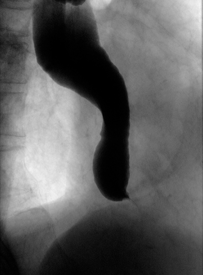 Fig. 1
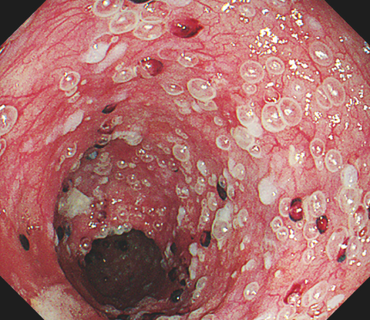 Fig. 2
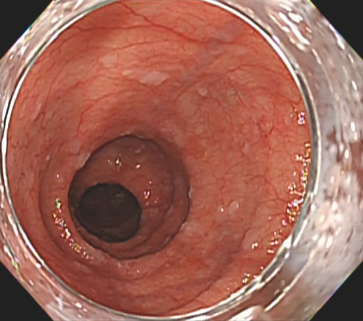 Fig. 3
Peer Reviews
No public reviews on file for this paper yet. If you reviewed it on a platform where reviews are public (OpenReview, ICLR, NeurIPS, ICML), you can paste yours below so the community can read it here.
Videos
No videos yet. Explain this paper in a talk, walkthrough, or lecture? Add one.
Taxonomy
TopicsAbdominal vascular conditions and treatments · Esophageal and GI Pathology · Eosinophilic Esophagitis
Although pneumatosis affecting the large intestine, small intestine, and stomach has been documented in the gastrointestinal tract, cases of pneumatosis in the esophagus are infrequent. The patient in this case was an octogenarian man who presented to Tokai University Hospital with the chief complaint of persistent dysphagia. Achalasia was diagnosed using upper gastrointestinal endoscopy and upper gastrointestinal imaging ( Fig. 1 ). Subsequently, hospitalization was deemed necessary due to compromised oral intake.
Upper gastrointestinal imaging revealed a smooth stricture, recognized as the "bird beak sign," featuring a maximum esophagus diameter of 3.9 cm and a flexion angle of 130°.
Upon admission, upper gastrointestinal endoscopy revealed conspicuous esophageal bubble lesions and esophageal blood clots ( Fig. 2 ). Conservative treatment measures were taken, and repeat upper gastrointestinal endoscopy was conducted to assess distinctive esophageal findings. Strikingly, all previously identified characteristic esophageal lesions had disappeared ( Fig. 3 ). Testing for viruses, autoimmune diseases, and drugs yielded negative results, and the patient was diagnosed with esophageal emphysema and blood clots caused by achalasia ( Video 1 ).
Endoscopy showed esophageal emphysematous changes and the presence of blood clots.
All previously identified characteristic esophageal lesions disappeared.
An unusual case of esophageal pneumatosis and hematomatosis associated with achalasia.Video 1
Gastrointestinal emphysema may manifest as either idiopathic or secondary, with idiopathic cases constituting 15% of all occurrences and secondary cases accounting for the remaining 85% 1 . The etiologies of secondary cases include necrotizing enterocolitis, pyloric stenosis, peptic ulcer disease, jejunoileal bypass, and intestinal obstruction. The mechanism underlying emphysema remains elusive, although the four proposed mechanisms include bacterial involvement, mechanical factors such as increased intraluminal pressure, mucosal damage allowing air entry, and lung disease-related air dislodgment 2 3 4 5 .
In this specific instance, the heightened gastrointestinal lumen pressure resulting from achalasia was postulated to penetrate the gastrointestinal wall through mucosal lacerations and invading small vessels, leading to esophageal mucosal emphysema and blood clot formation. Vomiting, a symptomatic manifestation, is believed to have contributed to esophageal emphysema via the entry of air from the esophagus into mucosal tears induced by mechanical irritation.
Endoscopy_UCTN_Code_CCL_1AB_2AC_3AH
The reference list from the paper itself. Each links out to its DOI / PubMed record.
- 1Greenstein AJ Nguyen SQ Berlin A Pneumatosis intestinalis in adults: management, surgical indications, and risk factors for mortality J Gastrointest Surg 2007111268127410.1007/s 11605-007-0241-917687617 · doi ↗ · pubmed ↗
- 2Shinagare AB Howard SA Krajewski KM Pneumatosis intestinalis and bowel perforation associated with molecular targeted therapy: an emerging problem and the role of radiologists in its management AJR Am J Roentgenol 20121991259126523169717 10.2214/AJR.12.8782 · doi ↗ · pubmed ↗
- 3Khalil PN Huber-Wagner S Ladurner R Natural history, clinical pattern, and surgical considerations of pneumatosis intestinalis Eur J Med Res 20091423123910.1186/2047-783x-14-6-23119541582 PMC 3352014 · doi ↗ · pubmed ↗
- 4Ho LM Paulson EK Thompson WM Pneumatosis intestinalis in the adult: benign to life-threatening causes AJR Am J Roentgenol 20071881604161310.2214/AJR.06.130917515383 · doi ↗ · pubmed ↗
- 5Blair HA Baker R Albazaz R Pneumatosis intestinalis an increasingly common radiological finding, benign or life-threatening? A case series BMJ Case Rep 20152015 bcr 201420723410.1136/bcr-2014-207234 PMC 433688425694632 · doi ↗ · pubmed ↗