The “echo-free space” technique: A safe and reliable method for endoscopic ultrasound scope insertion into the esophagus
Shunsuke Omoto, Mamoru Takenaka, Kota Takashima, Yoriaki Komeda, Masatoshi Kudo

Abstract
Genes, proteins, chemicals, diseases, species, mutations and cell lines named across the full text — each resolved to its canonical identifier and authoritative record.
Click any figure to enlarge with its caption.
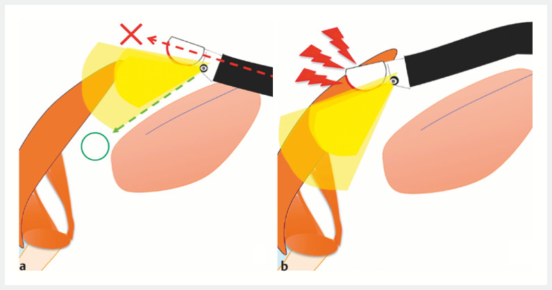 Fig. 1
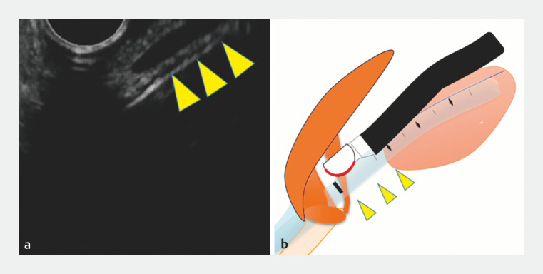 Fig. 2
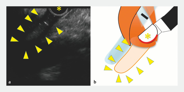 Fig. 3
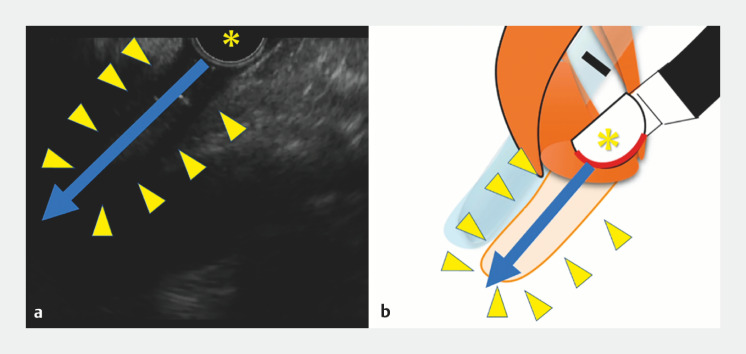 Fig. 4
- —Japan Society for the Promotion of Science10.13039/501100001691
Peer Reviews
No public reviews on file for this paper yet. If you reviewed it on a platform where reviews are public (OpenReview, ICLR, NeurIPS, ICML), you can paste yours below so the community can read it here.
Videos
No videos yet. Explain this paper in a talk, walkthrough, or lecture? Add one.
Taxonomy
TopicsEsophageal and GI Pathology · Gastroesophageal reflux and treatments · Esophageal Cancer Research and Treatment
In recent years, the use of endoscopic ultrasound-guided fine-needle aspiration (EUS-FNA) and EUS-guided drainage has greatly expanded 1 2 3 4 . However, inserting a linear EUS scope into the esophagus can be challenging, particularly in intubated or pediatric patients. This is because the scope is side-viewing, and the tip does not face the cervical esophagus. As a result, it can damage the laryngopharynx and lead to unsuccessful insertion if advanced, based on endoscopic imaging ( Fig. 1 ).
a The tip of the side-viewing scope does not face the cervical esophagus. b It can damage the laryngopharynx and lead to unsuccessful insertion if advanced, based on endoscopic imaging.
Because of these difficulties, inserting an EUS scope into the esophagus can be a hurdle for trainees learning this procedure. Even expert EUS sonographers may experience difficulties, resulting in repeated insertion attempts and potential damage to the laryngopharynx.
We present the case of an 8-year-old girl with an infected pancreatic pseudocyst who underwent emergency EUS-guided drainage under general anesthesia and tracheal intubation. Initially, scope insertion into the esophagus was difficult due to contact with the intubation tube ( Fig. 2 ). In this situation, the technique of inserting the EUS scope safely while confirming the lumen of the esophagus was required. In this case, we used the “echo-free space” technique, which is a safe insertion technique for EUS scopes that depicts the digestive tract lumen as echo-free space 5 .
The insertion of the scope into the esophagus was difficult due to contact with the intubation tube (arrowhead).
First, the cervical esophageal lumen, which is depicted by EUS as an echo-free space, was sought and successfully identified ( Fig. 3 ). Then, the tip of the scope was advanced toward the echo-free space ( Fig. 4 ). By aligning the tip of the scope with the echo-free space on the EUS image, successful insertion of the EUS scope into the esophagus was achieved ( Video 1 ).
The cervical esophageal lumen, which was depicted by EUS as an "echo-free space," was successfully identified (arrowhead).
The tip of the scope () was advanced toward echo-free space (arrowhead). By aligning the tip of the scope with the echo-free space on the EUS image, successful insertion of the EUS scope into the esophagus was achieved.*
This video introduces the “echo-free space” method, which allows safe insertion of the EUS scope while confirming the esophageal lumen as the echo-free space.Video 1
This echo-free space technique, which is the safe EUS scope insertion technique guided by an EUS image, can be helpful for trainees and experts when esophageal insertion of the EUS scope is difficult.
Endoscopy_UCTN_Code_TTT_1AS_2AB
The reference list from the paper itself. Each links out to its DOI / PubMed record.
- 1Takenaka M Okabe Y Kudo M Hemorrhage from metastasis of a 5-mm renal cell carcinoma lesion to the gallbladder detected by contrast-enhanced endoscopic ultrasonography Dig Liver Dis 20195174330545796 10.1016/j.dld.2018.11.010 · doi ↗ · pubmed ↗
- 2Takenaka M Omoto S Kudo M Endoscopic ultrasound fine-needle biopsy may contribute to the diagnosis of malignant lymph nodes Clin Endosc 20205350850910.5946/ce.2020.19932967410 PMC 7548144 · doi ↗ · pubmed ↗
- 3Tanaka T Omoto S Takenaka M Urgent endoscopic ultrasound-guided choledochoduodenostomy for adenocarcinoma of the ampulla of Vater with scirrhous invasion Dig Endosc 202133 e 43e 4410.1111/den.1391933506566 · doi ↗ · pubmed ↗
- 4van der Merwe S Wvan Wanrooij RLJ Bronswijk M Therapeutic endoscopic ultrasound: European Society of Gastrointestinal Endoscopy (ESGE) Guideline Endoscopy 20225418520510.1055/a-1717-139134937098 · doi ↗ · pubmed ↗
- 5Omoto S Takenaka M Maluf-Filho FA novel and effective EUS training program that enables visualization of the learning curve: Educational Program of Kindai system (EPOK)Video GIE 2022716516835585898 10.1016/j.vgie.2022.01.014PMC 9108189 · doi ↗ · pubmed ↗