Clinical Images in Emergency Medicine: Cushing’s Disease
Jason D. Vadhan, Nathaniel Hansen, Fernando L. Benitez, Larissa I. Velez

TL;DR
A young woman with Cushing’s disease was diagnosed after showing symptoms like weight gain and facial swelling, leading to discovery of a pituitary tumor.
Contribution
Highlights the importance of recognizing rare Cushing’s disease in emergency settings to avoid diagnostic delays.
Findings
A 22-year-old female was diagnosed with Cushing’s disease via MRI revealing a pituitary macroadenoma.
Symptoms like fatigue and weight gain can mimic common disorders, leading to delayed diagnosis.
Emergency physicians should maintain a high suspicion for Cushing’s disease in similar presentations.
Abstract
A 22-year-old female presented to the emergency department with a two-month history of worsening fatigue, unintentional weight gain, and progressive facial swelling. Physical examination findings included hirsutism, moon facies, and abdominal striae. Subsequent brain magnetic resonance imaging revealed the presence of a 2.4-centimeter pituitary macroadenoma, confirming the diagnosis of Cushing’s disease. The patient was then admitted for neurosurgical tumor resection. Cushing’s disease is exceedingly rare and often presents with symptoms resembling more prevalent disorders, contributing to delays in diagnosis. Therefore, maintaining a high index of suspicion for this disease is crucial for emergency physicians.
Genes, proteins, chemicals, diseases, species, mutations and cell lines named across the full text — each resolved to its canonical identifier and authoritative record.
Click any figure to enlarge with its caption.
 Image 1
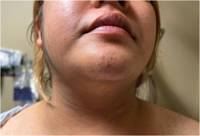 Image 2
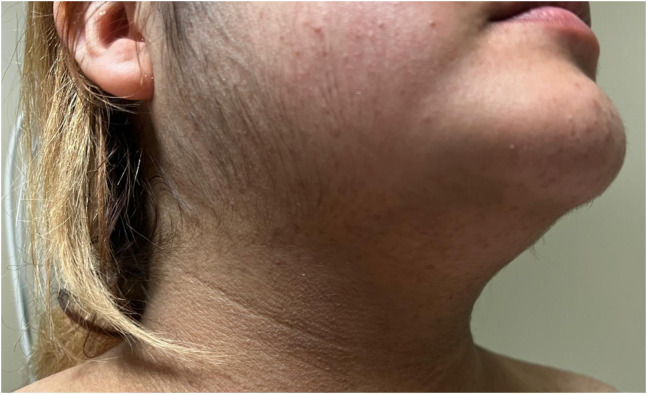 Image 3
Peer Reviews
No public reviews on file for this paper yet. If you reviewed it on a platform where reviews are public (OpenReview, ICLR, NeurIPS, ICML), you can paste yours below so the community can read it here.
Videos
No videos yet. Explain this paper in a talk, walkthrough, or lecture? Add one.
Taxonomy
TopicsPituitary Gland Disorders and Treatments · Adrenal and Paraganglionic Tumors · Glioma Diagnosis and Treatment
CASE PRESENTATION
A 22-year-old female with a past medical history of hypertension and diabetes presented to the emergency department with two months of abdominal striae, persistent fatigue, unintentional weight gain exceeding 30 pounds, and progressive facial swelling. Physical exam revealed the presence of abdominal striae (Image 1), facial and trapezius adiposity (Image 2), and hirsutism (Image 3). Since the patient was not receiving steroid therapy at the time, her symptoms raised suspicion for Cushing’s disease. Subsequently, a brain magnetic resonance imaging (MRI) with intravenous contrast was performed, revealing a 2.4-centimeter pituitary macroadenoma causing severe upward displacement of the optic chiasm (Supplementary Image). Neurosurgery was sought, and the patient was admitted for operative management.
Lower abdominal striae due to hypercortisolism.
Facial and trapezius adiposity, colloquially referred to as “moon facies” and a “buffalo hump.”
Hirsutism of the face due to increased adrenocorticotrophic hormone production, resulting in hyperandrogenism.
DISCUSSION
Cushing’s disease is a rare disorder characterized by excessive cortisol production from the adrenal glands, which can either be from the adrenals directly or from corticotropin-releasing tumors in the lungs or pituitary gland.1 The term “Cushing’s disease” refers explicitly to the presentation of Cushing’s syndrome caused by a pituitary tumor. Cushing’s disease is more commonly observed among women, typically appearing between 20–40 years of age.1 Clinical manifestations are attributed to increased cortisol production, which causes weight gain, fatigue, poor concentration, hypertension, hyperglycemia, excess hair growth, abdominal striae, adipose deposition, and menstrual irregularity.2 Unfortunately, these symptoms are nonspecific and overlap with common medical conditions such as diabetes, hypertension, and polycystic ovarian syndrome. Consequently, the diagnosis of Cushing’s disease is often delayed, with an average time to diagnosis exceeding three years from symptom onset.3
The evaluation for Cushing’s disease is typically initiated in an outpatient setting and involves various tests, including midnight salivary cortisol measurement, low-dose dexamethasone suppression test, or 24-hour urine-free cortisol level assessment.2 However, in the emergency department, obtaining a brain MRI may be warranted to detect a visible pituitary tumor, which can be seen approximately 50% of the time, as in this case.2 When pituitary tumors are discovered, neurosurgical consultation and operative resection are often necessary.
Supplementary Information
The reference list from the paper itself. Each links out to its DOI / PubMed record.
- 1Gardner DG Shoback DM . Greenspan’s Basic and Clinical Endocrinology. New York, NY: Mc Graw-Hill Education; 2017.
- 2Nieman L Swearingen B . Cushing’s syndrome and Cushing’s disease. CMM Global. 2013:1–12.
- 3Rubinstein G Osswald A Hoster E et al . Time to diagnosis in Cushing’s syndrome: a meta-analysis based on 5367 patients. J Clin Endocrinol Metab. 2019;105(3):e 12–22.10.1210/clinem/dgz 13631665382 · doi ↗ · pubmed ↗